Numerical chromosome abnormalities
Numerical chromosome abnormalities are deviations from the normal number of chromosomes. These deviations can refer to individual chromosomes, both in the sense of "plus" (supernumerary chromosome, extranumerary chromosome) and in the sense of "minus" (missing chromosome, missing chromosomes). The entire haploid set of chromosomes may also be multiplied. Unlike structural chromosome aberrations, chromosomes are not structurally altered in numerical aberrations. A number of numerical abnormalities are incompatible with life (all autosomal monosomies, most trisomies, polyploidy) and those that allow survival usually cause severe syndromes.
Aneuploidies[edit | edit source]
Aneuploidies are such changes in the number of chromosomes where there is one extra chromosome, i.e. trisomy (2n+1), or 1 chromosome is missing - monosomy (2n-1).
Trisomy[edit | edit source]
Trisomies are caused by nondisjunction, i.e. an error in the separation of homologous chromosomes in the 1st meiotic division, or chromatides in the 2nd meiotic division. The risk of this error, i.e. the occurrence of trisomy, depends significantly on the age of the mother, at the age of 25 the woman is roughly twice the population risk and then increases dramatically with age. The risk of this error is associated with the fact that meiotic division in a woman begins already during embryonic development, then is stopped and continues only at the time of sexual maturity. The age of fathers plays a role only from 50 years.
If errors in the number of chromosomes occur during meiotic division and an abnormal gamete is fertilized (in the case of nondisjunction, a disomic gamete is formed - it has an extra chromosome and a nullisomic gamete - it does not have the given chromosome), the error then occurs in all cells of the individual. If the error occurs postzygotic during the mitotic division of the zygote, a so-called mosaic occurs, where two or more cell lines with different karyotypes are present.
Most autosomal trisomies do not allow survival, only an individual can be born with trisomy 21, 18, 13, patients with trisomy 21 survive to adulthood. One more trisomy allows survival, trisome 8, but it always occurs in a mosaic with a predominance of the normal celly line i live births.
Monosomy[edit | edit source]
Monosomies arise from nondisjunction and fertilization of an abnormal - nullisomic gamete. Another mechanism by which only monosomy arises is the delay of the chromosome in anaphase and its non-incorporation into the daughter nucleus. Both mechanisms can occur in meiosis, or postzygotic during mitotic division of the zygote. The only monosomy that allows survival is monosomy X, monosomy of autosomes we would only find in spontaneous abortions.
Polyploidy[edit | edit source]
Polyploidy is the multiplication of the chromosomal set, in triploidy there are 69 chromosomes, i.e. three time the haploid ste (3n), in tetraploidy 92 chromosomes - four times the haploid set (4n).
Triploidy[edit | edit source]
Triploidy is a lethal genetic constitution, rarely an individual is born with triploidy but dies soon after birth. Triploidy is most often caused by a fertilization disorder, so-called dispermia (fertilization of an egg by two sperm), less often it is the result of the fusion of an abnormal unreduced gamete (created by nondisjunction of all chromosomal pairs) and a normal gamete. The phenotype of the abnormal product depends on whether the supernumerary set of chromosomes is of paternal origin (partial moles) or maternal origin (nonmolar product). This situation (a different phenotype depending on the parental origin of the supernumerary set of chromosomes) is caused by gene imprinting, which manifests itself at the beginning of embryonic development (active paternal alleles of certain genes are responsible for the development of the membranes, active maternal alleles of other genes for the development of the embryo itself). if the mutual function of the imprinted genes is not balanced, as is the case with triploidy, for example, a pathological product is produced.
Tetraploidy[edit | edit source]
Tetraploidy is a lethal genetic constitution where the number of chromosomes in an individual is 92 (4n). Tetraploidy results from endoreduplication, i.e. division of chromosomes, without cell division.
Numerical aberrations of autosomes[edit | edit source]
Down's syndrome[edit | edit source]
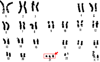
Down's syndrome (DS) is one of the most well-known syndromes caused by a chromosomal abnormality.
It is the most common syndrome caused by trisomy of a chromosome in live births (specifically trisomy of chromosome 21) and the most common congenital cause of mental retardation. Other characteristic clinical symptoms are congenital heart defects, muscle hypotonia and a typical appearance (eyes with a skin fold - epicantherum - and upward-facing eye slits), as well as a monkey furrow, clinodactyly of the 5th finger, etc. In newborns, a large, crawling tongue is noticeable. In addition to greater susceptibility to infections, they also have an increased risk of cancer, especially leukemia. This is due to the fact that trisomic cells are much more sensitive to the effect of mutagnes and carcinogens. Although this is impairment allows survival, a large proportion (about ¾) of all resulting trisomies 21 are aborted.
In about 4 % of patients born with Down syndrome, the third, the redundant chromosome 21 is translocated to one of the acrocentric chromosomes (the so-called translocation form of trisome caused by Robertsonian translocation). While the risk of free trisomy depends on the age of the mother (nondisjunction risk), the risk of the translocation form of trisomy does not depend on the age of the parents, but in the case of the translocation form, in about half of the cases one of the parents is a carrier of the balanced form of the translocation and there is a risk for the offspring who may inherit this translocation in unbalanced form. If the parent is a carrier of the so-called homologous fusion of both 21 st chromosomes, the risk of DS or miscarriage is even 100 %. In non-homologous Robertsonian fusion, the risk is considerable lower, but carrying a balanced aberration is always a reason for prenatal cytogenetic diagnosis.
A small percenatge of DS patients have a mosaic form of trisomy, resulting from postzygotic loss of chromosome 21 from a trisomic zygote or postyzygotic nondisjunction during division of a normal zygote.
Thanks to modern methods of prenatal diagnosis, this syndrome can be diagnosed in the overwhelming majority of cases already during pregnancy. Indications for prenatal cytogenetic diagnosis, which lead to the detection of numerical chromosomal changes, are older age of the mother, atypical biochemical screening, abnormality on ultrasound, including minor morphological deviations. Of course, carrying a balanced Roberstonian translocation is also an indication for this examination.
Edwards syndrome[edit | edit source]
Edwards syndrome (ES) is caused by trisomy od 18th chromosome. j
From the clinical symptoms, there is a noticeable flexion deformity of the fingers (the index finger and the 5th finger overlap the other fingers), atypical facies with micrognathia, prominent head, severe growth retardation, congenital defects of the heart and kidneys, hypotonia.
The average length of survival is about 2 moths, quite exceptionally the patient survives 1 year, but the vas majority of all resulting trisomies 18 are aborted.
Patau syndrome[edit | edit source]
Patau syndrome (PS) is cause by trisomy 13.
Manifestations include microcephaly, sloping forehead, hypertelorism, microphthalmia and even anophthalmi or cyclopia, cleft lip and palate, micrognathia, may be polydactyly. Those affected have congenital developmental defects of the brain (holoprosencephaly), heart defects and defects of other organs.
Affected people usually die within the first month of life. A translocation form of trisomy 13 is also known. The majority of all resulting trisomies 13 are aborted.
Numerical aberrations of gonosomes[edit | edit source]
Turner syndrome[edit | edit source]
Turner syndrome (TS) is most often caused by monosomy of the X chromosome (karyotype 45,X), or by various structural aberrations (for example, a deletion on the X chromosome, etc.). Numerical and structural changes can also occur in a mosaic. Classic manifestations are: short stature, ovarian dysgenesis associated with sterility, in some patients there is a skin fold (pterygium colli). Intelect is not impaired. In newborns, there is early lymphedema on the limbs, which should signal the suspicion of TS and the indication of cytogenetic examination. Phenotype modification with growth hormone and hormone replacement therapy is possible.
In the case of a structural aberration, the so-called isochromosome for the long arms of the X chromosome, the patient's phenotype corresponds to TS, but the patient can be fertile, as in the case of deletion of the short arms of the X chromosome. If the so-called critical region on the long arms of the X chromosome is preserved, fertility is preserved. Conversely, a deletion or a break in this critical region (when an X / autosomal translocation occurs) leads to gonadal dysgenesis, even if the patient's phenotype does not correspond to TS. Mosaics are very common in TS, when a chromosome is lost, not only the X is loste, but there can also be a very early loss of the Y chromosome (the paternal gonosome is more often lost). If the mosaic is 45,X/46,XY, there is a risk of malignancy of the gonad.
It is interesting that up to 99 % of all resulting X monosomies are aborted. It is assumed that thoso monosomies that survive are actually mosaics, although often diagnosed as full X monosomies.
Klinefelter syndrome[edit | edit source]
Klinefelter syndrome (KS) results from the presence of an extra X chromosome in a male. It is most often caused by karyotype 47,XXY, variants with more X chromosomes (48,XXXY or 49,XXXXY) are also possible, which have a more pronounced manifestation. There are also mosaic forms. The main symptoms are: infertility (azoospermia), hypogonadism, average to tall height, long limbs, sparse hair, gynecomastia. In the case of the 49,XXXXY karyotype, the patient is retarded and the phenotype resembles Down syndrome, with the difference that the KS patient is tall, unlike the DS patient.
Syndrome 47,XXX[edit | edit source]
X chromosome trisomy, also called three X syndrome (and formerly "Superfemale syndrome"). As the name already suggests - it is caused by kayrotype 47,XXX, it can also occur in mosaic. Karyotype 48,XXXX or 49,XXXXX can also occur very rarely, these cases have a different and more pronounced manifestation. The 47,XXX syndrome itself does not have a distinct clinical picture, come women are examined for infertility. Reduced fertility would be in the case of mosaic 45,X/47,XXX. Otherwise, a 47,XXX woman may be fertile, but some of her gametes may be abnormal. Minor problems of a psychosocial nature may occur, for example learning problems and an increased tendency to schizophrenia is also reported.
Syndrome 47,XYY[edit | edit source]
This syndrome is caused by the presence of two or more Y chromosomes in the karyotype, most often the karyotype 47,XYY. Previously, this syndrome was referred to as "Supermale" - a term that is no longer used today. Males can be taller, it was previously thought that males with two Ys were more aggresive,but this has not been confirmed. There are completely normal men in the population with two Ys in their cells.
Links[edit | edit source]
Related articles[edit | edit source]
- Chromosomal abnormalities
- Chromosome aberrations in the etiology of neoplasia
- Tumor cytogenetics
- Chromosomal mosaicism
References[edit | edit source]
- PRITCHARD, Dorian J. – KORF, Bruce R.. Základy lékařské genetiky. 1.. edition. Galén, 2007. 182 s. pp. ISBN 978-80-7262-449-2.
- NUSSBAUM, R. L. – MCINNES, R. R. – WILLARD, H. W.. Klinická genetika (Thompson&Thompson). 6. edition. Triton, 2004. ISBN 80-7254-475-6.
- ŠÍPEK, Antonín. Genetika [online]. [cit. 30. 5. 2009]. <http://www.genetika-biologie.cz/chromozomove-aberace>.


