Oesophagus - structure (macro and micro), syntopy
Oesophagus - Structure (Macro and Micro), Syntopy
Introduction
The oesophagus is a muscular tube that serves as the conduit for food and liquids from the pharynx to the stomach. Its structure and relationships (syntopy) are crucial for understanding its role in both normal physiology and pathological conditions.
Macroscopic Structure
Length and Location:
- The oesophagus is approximately 25 cm long in adults, extending from the level of the cricoid cartilage (C6 vertebra) to the gastroesophageal junction (T11 vertebra).
- It passes through the neck, thoracic cavity, and diaphragm before entering the abdominal cavity.
Regions:
- Cervical oesophagus: Begins at the cricopharyngeus muscle.
- Thoracic oesophagus: Extends through the superior and posterior mediastinum.
- Abdominal oesophagus: Approximately 1-2 cm, terminating at the cardiac orifice of the stomach.
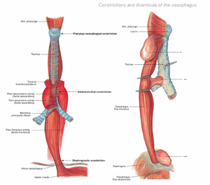
Constrictions: The oesophagus has three natural constrictions:
- At the pharyngoesophageal junction (15 cm from incisors).
- Where it is crossed by the aortic arch and left main bronchus (22.5 cm and 27.5 cm, respectively).
- At the oesophageal hiatus in the diaphragm (40 cm from incisors).
Layers:
- Adventitia/Serosa:
- Cervical and thoracic oesophagus have an outer adventitial layer.
- The abdominal oesophagus is covered by serosa.
- Muscularis propria:
- Upper third: striated muscle.
- Middle third: mixed striated and smooth muscle.
- Lower third: smooth muscle.
- Submucosa: Contains esophageal glands secreting mucus.
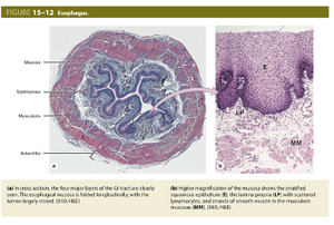
- Mucosa: Lined with non-keratinized stratified squamous epithelium for protection against friction.
Microscopic Structure
Epithelium:
- The mucosa is composed of non-keratinized stratified squamous epithelium.
- Transition to columnar epithelium occurs at the gastroesophageal junction (Z-line).
Lamina Propria:
- Loose connective tissue supports the epithelium and contains lymphatic tissue.
Muscularis Mucosae:
- Thin layer of smooth muscle that aids in motility.
Submucosa:
- Dense connective tissue housing esophageal glands and blood vessels.
- Meissner’s plexus is present.
Muscularis Propria:
- Inner circular and outer longitudinal layers.
- Auerbach’s (myenteric) plexus lies between these layers.
Adventitia/Serosa:
- Composed of connective tissue.
- Provides structural support and flexibility.
Syntopy -
Cervical Region:
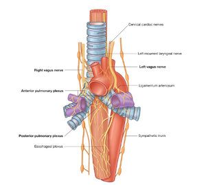
- Anterior: Trachea and recurrent laryngeal nerves.
- Posterior: Vertebral column.
- Lateral: Common carotid arteries and thyroid lobes.
Thoracic Region:
- Anterior: Trachea, left bronchus, and pericardium.
- Posterior: Thoracic vertebrae and thoracic duct.
- Right: Azygos vein and pleura.
- Left: Aortic arch, left subclavian artery, and thoracic aorta.
Abdominal Region:
- Anterior: Left lobe of the liver.
- Posterior: Left crus of the diaphragm.
Sources
- Gray’s Anatomy for Students, 4th Edition.
- Sobotta Atlas of Human Anatomy, 16th Edition.


