Organ of Corti
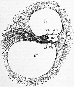
The organ of Corti is the key unit of sound perception, it generates a nerve impulse from the mechanical energy of sound waves. It is located on the lamina basilaris in the ductus cochlearis of the cochlea of the inner ear. It is composed of both secondary sensory cells (hair cells) and support cells.
The outer hair cells (about 20,000) form 3-4 rows with the function of amplifying sounds. The inner hair cells (about 4,000) with the function of their own receptor are arranged in one row. There are several types of support cells, differing in shape, function and location. They include Hensen's cells, Deiters' cells (phalangeal), Claudius' cells, Boettcher's cells, cells of the outer and inner Corti's pillar, which form a so-called triangle of Corti between the outer and inner hair cells.
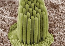
Stereocilia protrude from the apical part of the sensory cells and are covered by the so-called tectorial membrane, a gel-like substance rich in glycoproteins. Irritation of the hair cells occurs when the stereocilia are irritated by movement of the tectorial membrane. This movement results from the transmission of sound waves from the external environment through the eardrum, auditory ossicles, oval window, perilymph, and endolymph just to the tectorial membrane (or bone conduction). The bending of the stereocilia evokes a receptor potential by depolarization, which is further propagated through the auditory portion of the fibers of the VIII. cranial nerve.
Schematic of the organ of Corti in the blanitic labyrinth[edit | edit source]
- 1 – perilymph
- 2 – endolymph
- 3 – tectorial membrane
- 4 – The organ of Corti
- 5 – inner hair cells
- 6 – outer hair cells
- 7 – inner Corti pillar
- 8 – outer Corti pillar
- 9 – Deiters' cells (phalangeal)
- 10 – Hensen cells (outer border)
- 11 – Claudius cells (transition into the sulcus spiralis externus)
- 12 – basilar membrane
- 13 – Ductus cochlearis
- 14 – triangle of Corti
- 15 – sulcus spiralis internus
- 16 – scala tympani
- 17 – lamina spiralis ossea
- 18 – auditory nerve fibres
- 19 – efferent fibres
- 20 – afferent fibres
Links[edit | edit source]
Related articles[edit | edit source]
External links[edit | edit source]
Sources[edit | edit source]
- HYBÁŠEK, Ivan – VOKURKA, Jan. Otorinolaryngologie. 2. edition. Karolinum, 2006. 426 pp. ISBN 80-246-1019-1.
- VOKURKA, Martin – HUGO, Jan. Velký lékařský slovník [online] . 8. edition. Maxdorf, 2009. 1144 pp. Available from <http://lekarske.slovniky.cz/>. ISBN 978-80-7345-166-0.


