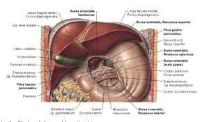
Parietal and Visceral Layers, Greater and Lesser Omentum
Parietal and Visceral Layers, Greater and Lesser Omentum
The peritoneum is a continuous, serous membrane lining the abdominal cavity and covering its organs.
It forms the largest serous membrane in the body and plays crucial roles in supporting abdominal organs and facilitating their movements.
Below is a detailed explanation based on Gray’s Anatomy and Sobotta Atlas of Human Anatomy.
1. Parietal and Visceral Layers
· Parietal Peritoneum:
o Lines the internal surface of the abdominal and pelvic walls.
o Innervated by somatic nerves (phrenic, lower intercostal, iliohypogastric, and ilioinguinal nerves), making it sensitive to pain, pressure, and temperature.
o Pain is localized to the specific dermatome corresponding to the innervation.
· Visceral Peritoneum:
o Covers the external surfaces of most abdominal organs.
o Innervated by autonomic nerves (sympathetic and parasympathetic fibers), making it sensitive to stretch and chemical irritation but not pain.
o Pain from visceral peritoneum is often referred to dermatomes associated with the affected organ.
Both layers secrete serous fluid, allowing frictionless movement of organs during digestion and respiration.
2. Greater and Lesser Omentum
· Greater Omentum:
o A large, apron-like fold of peritoneum that extends from the greater curvature of the stomach and proximal duodenum, draping over the intestines before attaching to the transverse colon.
o Composed of four layers of peritoneum.
o Functions:
o Fat storage: Contains adipose tissue for energy storage and insulation.
o Immunity: Contains lymphatic tissues and can adhere to inflamed areas, helping localize infections (e.g., “policeman of the abdomen”).
o Protection: Physically cushions abdominal organs.
· Lesser Omentum:
o A smaller fold of peritoneum connecting the lesser curvature of the stomach and proximal duodenum to the liver.
o Contains two peritoneal layers and consists of two parts:
§ Hepatogastric ligament: Extends between the liver and the stomach.
§ Hepatoduodenal ligament: Extends between the liver and duodenum, containing the portal triad (portal vein, hepatic artery, and bile duct).
o Functions:
o Provides a pathway for structures in the portal triad.
o Maintains the position of the stomach and liver.
3. Clinical Correlations
· Peritoneal Cavity:
o The space between the parietal and visceral layers. In males, it is closed; in females, it communicates with the external environment through the uterine tubes, uterus, and vagina.
o Excess fluid accumulation in the cavity is called ascites, commonly associated with liver disease or peritoneal infections (peritonitis).
· Omental Bursa (Lesser Sac):
o A potential space located posterior to the stomach and lesser omentum.
o Communicates with the greater sac through the epiploic foramen (of Winslow).
· Surgical Considerations:
o The greater omentum is often mobilized in surgeries to cover wounds or prevent adhesions.
o The hepatoduodenal ligament’s portal triad is a key structure in trauma and surgery, especially during procedures like the Pringle maneuver to control bleeding.
The peritoneum, with its parietal and visceral components and specialized structures like the greater and lesser omenta, plays a critical role in supporting abdominal viscera, enabling mobility, and mediating immune and metabolic functions. Its clinical relevance spans across surgery, infection, and abdominal pathology.

