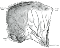
Parietal bone
The parietal bone is the paired bone of the cranial vault forming the top of the head. It has a square shape and is slightly arched by convexity. It consists of a single part, which is connected to the second parietal bone and to the other bones of the skull at the seams.
Anatomical Structures[edit | edit source]
Outer surface[edit | edit source]
- Similar to the frontal bone, it contains a curved tuber parietale' - the place where ossification of the parietal bone began.
- Linea temporalis superior - the upper raised bar for joining the fascia m. temporalis.
- Similarly, linea temporalis inferior - the lower bar for connecting the edge of the muscle.
- Sometimes the foramen parietale is visible as an opening for the venous sphincter.
Internal Area[edit | edit source]
It contains impressions of arteries brains (sulcus arteriae meningeae mediae) and venous vessels (sulcus sinus sagittalis superioris).
Connection of the parietal bone with the surroundings[edit | edit source]
The parietal bone is connected to the bones of the skull by these sutures:
- ventrally in the sutura coronalis' (crown suture) with os frontale;
- sagittally in the sutura sagittalis' (sagittal suture) with the second parietal bone;
- dorsally in sutura lambdoidea (lambda suture) with occipital bone;
- laterocaudally in sutura squamosa (scaly suture) with temporal bone.
Variation[edit | edit source]
This includes the os parietale bipartitum, when the ossification center does not merge and is divided into two, so the whole consists of two bones stacked on top of each other. Other variations include small bones in the sutures (os bregmatica, os preinterparietale) or different placements of the foramen parietale.
Links[edit | edit source]
Related links[edit | edit source]
References[edit | edit source]
- ČIHÁK, Radomír. Anatomy I. 2. edition. Prague : Grada, 2001. 516 pp. pp. 159-161. ISBN 978-80-7169-970-5.


