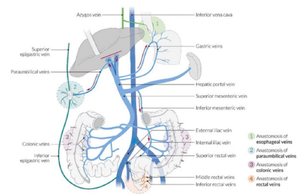
Portocaval Anastomosis
Portocaval Anastomosis[edit | edit source]
Porto-Caval Anastomosis - Collateral communication between the portal and the systemic venous system
- When there is a blockage of the portal system, portocaval anastomosis enable the blood to still reach the systemic venous circulation
- The importance of portocaval anastomoses is to provide alternative routes of circulation when there is a blockage in the liver or portal vein. These routes ensure that venous blood from the gastrointestinal tract still reaches the heart through the inferior vena cava without going through the liver
The major sites of these anastomoses include:[edit | edit source]
- Oesophageal – Between the oesophageal branch of the left gastric vein and the oesophageal tributaries to the azygous system
- Rectal – Between the superior rectal vein and the inferior rectal veins
- Retroperitoneal – Between the portal tributaries of the mesenteric veins and the retroperitoneal veins
- Paraumbilical – Between the portal veins of the liver and the veins of the anterior abdominal wall
Clinical significance[edit | edit source]
- If a large volume of blood passes through these anastomosis/connections, the veins become dilated resulting in:
- Oesophageal varices – oesophageal
- Haemorrhoids/piles – rectal
- Caput medusae – paraumbilical
References[edit | edit source]
- HUDÁK, Radovan – KACHLÍK, David. Memorix anatomie. 2. edition. Triton, 2013. ISBN 978-80-7387-712-5.


