Pregnancy
Physiological pregnancy can be described as the period from "implantation" of the fertilized egg into the lining of the uterus until "delivery" of the mature fetus. The average duration is set at 280 days = 40 completed weeks + 0 days = 10 lunar months. We call the duration after 40 weeks as "post-term pregnancy", which should not exceed 42+0. In such a case, we terminate the pregnancy by iatrogenic induction of labor.
Clinically, we divide pregnancy into three trimesters, according to the characteristic developmental changes for the given section (1. trimester to 12+6, 2. trimester to 27 +6 and 3rd trimester' until the date of birth).
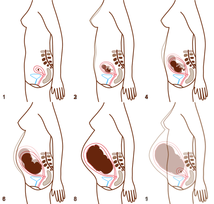
The course of pregnancy is associated not only with changes in the fetus, but also with physiological changes in the mother's body, which create ideal conditions for the development and growth of the fetus.
Detection of early pregnancy (pregnancy test)[edit | edit source]
Fertilization of the egg occurs during the period of "fertile days", which is on average about 3 days before and 3 days after ovulation (14th day of the cycle). However, this figure differs for each woman depending on the length of the cycle, menstruation, etc. Fertilization most often occurs in the tube area. The fertilized zygote then travels towards the uterine body, where it implants in the endometrium. The implanted embryo starts to produce the hormone hCG (chorionic gonadotropin), which ensures the stimulation of the corpus luteum (progesterone and estrogen production), which is necessary for the maintenance and development of pregnancy until the 5th-7th week. week (progesterone) and at the same time to induce physiological changes in the mother's body (estrogens).
The hCG values double every 2-3 days of pregnancy, the highest values are reached on the 90th day, from which they start to decrease again and stabilize over time and remain the same until the end of the pregnancy. Already in the first weeks of pregnancy, hCG values are detectable not only in the blood, but also in the urine. The non-invasive form of the test (dip dg. strips in urine) is a basic diagnostic examination for the detection of incipient pregnancy. We perform the test after a missed period. Sensitive tests can reliably diagnose an ongoing pregnancy as early as 14. day from implantation (values up to 500 IU/l), with values above 1500 IU/l pregnancy is evident even on ultrasound.
![]() If hCG values are above 1500 IU/l and we see an empty uterus on USG, it is probably an ectopic pregnancy.
If hCG values are above 1500 IU/l and we see an empty uterus on USG, it is probably an ectopic pregnancy.
Determining the gestational age of the fetus[edit | edit source]
In clinical practice, it is not possible to accurately determine the time of zygote implantation. We estimate the age of pregnancy based on knowing the date when the first day of the last menstrual period took place. We add 7 days to this date and subtract 3 months to determine the due date. We always make this estimate after the diagnosis of pregnancy, together with the assessment of the risk of the pregnant woman. The specific date of birth will allow us to better 'assess the developmental state of the fetus, given its gestational age.
Due to the not always accurate data on menstruation, today we prefer to re-evaluate the age of the fetus by ultrasound. We do this as part of screening in the 1st trimester, when we measure the CRL value (parietal-coccal distance), which we compare with standardized values and determine the gestational age of the fetus accordingly.
Physiological changes in the mother's body[edit | edit source]
First trimester[edit | edit source]
First to third week[edit | edit source]
After the fusion of the male and female gametes, the furrowing of the egg and the formation of a blastocyst begin. On the sixth day, the blastocyst is implanted in the uterus. The mother does not register this process in any way. In the second week, the embryo sinks deeper into the wall of the uterus, while light bleeding, which can be confused with menstruation, may occur. The third week is characterized by the establishment of the body axes of the future embryo. There is a blood connection between the mother and the fetus. As a result of hormonal changes, early signs of pregnancy are induced.
Embryonic stage[edit | edit source]
It lasts from the third to the eighth week. During this period, cell differentiation occurs, they establish and form all organ systems. During this period, the child is most susceptible to the action of external (teratogenic) factors that can cause subsequent developmental defects. Such factors include alcohol, drugs, toxins, infections, radiation, nutritional deficiency.
Fetal stage[edit | edit source]
At the beginning of the third month, the embryo turns into a fetus and the period of growth begins. The child grows in the amniotic cavity, takes on a human appearance and is connected to the mother's placenta. Until the end of the first trimester, pregnancy is accompanied by initial symptoms (see above).
Second trimester[edit | edit source]
In the fourth month, the rapid growth of the child continues, which is also reflected in the mother's weight gain. The symptoms of the first trimester will mostly disappear, but nausea, headaches, and tremors may persist, especially after eating. Glucose in the mother's blood rises according to the baby's needs, which causes the mother to feel energized. Hormonal changes are a sign of moodiness during this period. The most noticeable change during this period is the slowing down of head growth compared to the rest of the body. At the beginning of the fifth month, the length of the head corresponds to approximately one third of the occipital (TK) length. In the 5th month, the fetus grows very intensively, while the weight increases slowly. The surface of the body is covered with lanugo (short hairs), there are short hairs on the head and eyelashes on the eyelids. It is important that the fetus begins to move in the fifth month and the mother perceives these movements. A prematurely born fetus at the beginning of the 6th month hardly survives (respiratory system and CNS are not sufficiently developed). Between 6.5 and 7 months, it has a 90% chance of surviving outside the mother's body.
Third trimester[edit | edit source]
During further development, the weight of the fetus increases sharply. This is especially evident during the 2.5 months before childbirth, when up to 28 g is added per day. A woman's belly takes on the shape of a drop and the woman is able to move it up and down. The movements of the fetus are pronounced and sometimes disturbing for the woman. This period is also quite uncomfortable due to poor bladder control and back pain. Before birth, the fetus is covered with a substance - vernix caseosa, which consists of the secretion of sebaceous glands and desquamated cells of the epidermis. The measurements of the fetus just before the due date are: head circumference = 34 cm, weight = 3000–3400 g, BMI = 36 cm. However, these values are only approximate.
Birth[edit | edit source]
The date of birth is set approximately on the 266th day, i.e. the 38th week after conception. It represents the 280th day, i.e. the 40th week from the first day of the last period. A baby that is born prematurely is premature and a baby that is born later is considered to be transferred. Childbirth can be divided into three phases: 1st phase: opening, 2nd phase: the birth of the fetus itself, 3rd stage: delivery of the placenta and amniotic sac.
Postnatal period: begins immediately after birth and lasts about 6 weeks. During this period, the mother's body returns to its pre-pregnancy state. The uterus returns to its original size and the hormonal balance is balanced.
Links[edit | edit source]
References[edit | edit source]
References[edit | edit source]
- PAŘÍZEK, Antonín. Book About Pregnancy and Child. 3. edition. Galén, 2008. 752 pp. ISBN 9788072625949.
- ZDENĚK, Hájek. Obstetrics : 3rd, completely revised and supplemented edition. - edition. Grada Publishing, a.s., 2014. 1599 pp. ISBN 9788024745299.


