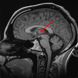
Prosencephalon
The proper prosencephalon comes into relevance at the end of the 4th week of embryonic development (28 days). At this point the cranial portion of the neural tube consists of prosencephalon (future telencephalon and diencephalon), mesencephalon (future midbrain), and rhombencephalon (future metencephalon and myelecephalon). The prosencephalon undergoes growth and proliferation and by the 8th week of embryonic development, splits into the telencephalon (future cerebral hemispheres) and diencephalon (pituitary, hypothalamus, thalamus, pineal gland, and optic cup).
Derivatives of the Prosencephalon[edit | edit source]
The diencephalon is a structure of great importance when it comes to establishment of sensory pathways to the cortex and future memory centers. The alar plates form the medial walls of future thalamus and hypothalamus (thus future 3rd ventricle lateral walls). The hypothalamic sulcus splits this plate into upcoming thalamus and hypothalamus. Pineal gland is formed by an evagination of the caudal end of diencephalon and will later join the upper limits of the adult midbrain. The posterior part of the pituitary gland (pars nervosa) is formed from the diencephalic projection know as the infundibulum. The anterior part of the pituitary (pars distalis) is formed from an invagination anterior to the oropharyngeal membrane, known as Rathke's pouch. These two pituitary regions meet at an intersection known as pars intermedia.
The telencephalon leads to the formation of the cerebral hemispheres, centers of conscious motoric motion, forethought, sensory perception, and many others. The hemispheres begin as two pockets on either side of the main telencephalic column joined by the lamina terminalis in a medial position (this lamina is also the most cephalic portion in the early CN system). The corpus striatum (name derived from histological appearance) forms within the telencephalon and then splits into the caudate nucleus and lentiform nucleus. This split is denoted in the adult brain by the internal capsule which contains mostly efferent motoric fibers from the future cortex. The telencephalon continues to differentiate and form the trademark gyri of the cortex. This expansion happens around the telencephalon (except in the innermost part of the future lateral fissure, called the insula) and gives rise to the frontal, parietal, temporal and occipital lobes.
Links[edit | edit source]
Related Articles[edit | edit source]
Bibliography[edit | edit source]
SADLER, W. T – LANGMANN, J. Langman's Medical Embryology. 12th edition. Philadelphia : Wolters Kluwer Health/Lippincott Williams & Wilkins, 2012. ISBN 9781451113426.
DUDEK, Ronald W – FIX, James D. Embryology. 6th edition. Philadelphia : Wolters Kluwer Health/Lippincott Williams & Wilkins, 2011. ISBN 9781605479019.


