Renal colic
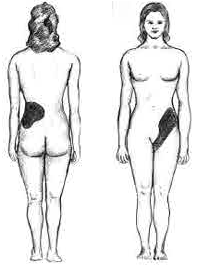
Renal colic is pain of variable intensity, usually spreading from the hips to the lower abdomen or genitals (but also vice versa). It originates in the kidney or upper urinary tract, and is caused by obstruction of the urinary tract.
Obstruction can be caused by one of the following reasons:
- Urolithiasis – concrete stuck on the level of pyeloureteral junction, ureter, ureterovesical junction,
- Blood clot passage hematuria,
- Passege of tumor or necrotic masses (TBC, necrotic papilitis),
- Passage of pus (pyelonephritis).
Characteristic[edit | edit source]
- Pain of varying intensity which, shoots to:
- to the side and back (origin in the upper part of the urinary tract),
- to scrotum (labia) and medial areas of the thigh( origin in the lower urinary tract),
- vegative accompaniment ( from sweating, nauzea, vomitng) to paralytic ileus,
- motoric disturbance (finding a relief position for the patient),
- hematuria (micro- i macroscopic) (is rarely missing and it is in cases with complete obstruction of the ureter by concrete),
- polacisuria – in intramural bladder calculus.
Diagnostic - examination sequence[edit | edit source]
- Anamnesis – clinical symptoms
- Physical examination – palpation pain in the urethra, positive bimanual palpation of the kidney and tapottment on the affected side
- Chemical examination of urine + sedimentation – higher erythrocytes and leucocytes , sometimes a crumb with crystals (oxalates, urates)
- USG – on ultrasound dilatation of the hollow system of the kidney on the affected side, or a hyperechogenic signal of the concretion, if there is a pyeloureteral or ureterovesical junction in the area.
- CT – in case of a positive finding on USG we will perform native CT of the kidneys for more accurate localisation of the concrete
- Native nephrogramy - alternative examination to CT.
- Imaging methods such as excretory urography and ascending / antegrade ureteropyelography are less widely used today.
Diferencial diagnostic[edit | edit source]
- A sudden abdominal event (biliar colic, ileus, apendicitis, ovarian torsion)
- Adnexitis (inflammation of the fallopian tube)
- Acute pyelonephritis (it can be also characterized by acute pain in the hips, it can be without temperatures)
Treatment[edit | edit source]
The goal of renal colic treatment is to release obstruction in the urine outflow, including maintaining the morphology and function of the kidney and urinary tract.
- Konservative – expelling – with a concretion diameter of up to approximately 4-5 mm, when we expect spontaneous concretion departure (effectiveness cca 50 %[1])
- spasmolytics, α-blocators (tamsolusin), antiphlogistics – to relieve of the ureteral edema (indometacin, diklofenak, escin), analgetics (Algifen)
- infusion of the Hartmann solution with Algifenem
- Ureterorenoscopy (URS) – extraction of the concrete by Dorma basket or ticks, method of choice for concretions > 5 mm wedged in the distal ureter area. For larger stones, there is a possibility of crushing with a contact lithotripter with subsequent extraction of fragments.
- Acute derivation of urine
- If it is not possible to extract the concrete, an ureteral stent is inserted for bypassing the obstruction
- In the risk of urosepsis ( colic with high temperatures) or renal insufficiency, urine drainage by the path nephrostomy
- Another methods for removing ureterolithiasis
- Extracorporal litotrypsy by shock wave – suitable for stones located in the proximal section of the ureter and thus localisable ultrasonographically. The advantage is that the patient doesn't have to be in general anesthesia.
- Ureterolithotomy – choice for large urethral-wedged stones
Following care[edit | edit source]
After the concrete removing, it is neccessary to control the patient condition – ultrasonic exclusion of congestion in hollow system, native nephrogramy to verify the right position of the inserted stent. Prolonged blockade of ureterolithiasis can lead to renal dysfunction with subsequent nephrectomy.
To prevent recurrence metaphylaxis must be also initiated, for example patient should be inform about strict and adequate hydratation, exercise, reduce a risky groceries (low purine diet for an urate lithiasis - reduce consummation of the sea-food).
Links[edit | edit source]
Related articles[edit | edit source]
Used literature[edit | edit source]
- KAWACIUK, Ivan. Urologie. 1. edition. Galén, c2009. ISBN 978-80-7262-627-7.
- HERÁČEK, Jiří – URBAN, Michael. Urologie pro studenty [online] . 2.0. edition. 2014. Available from <http://www.urologieprostudenty.cz>. ISBN 978-80-254-1859-8.
- PASTOR, Jan. Langenbeck's medical web page [online]. [cit. 3.6.2010]. <http:https://langenbeck.webs.com/>.
Reference[edit | edit source]
- ↑ KAWACIUK, Ivan. Urologie. 1. edition. Galén, c2009. ISBN 978-80-7262-627-7.

