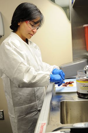
Sample processing for histological examination
Sample fixation[edit | edit source]
After the tissue sample is taken is taken, the regulatory relationships of the cells, the oxygen supply and the disorganization of the cellular metabolism will be interrupted. The activity of the cell's own enzymes causes autolysis (postmortem self digestion) of the cells. Microorganisms can also spoil the sample (rotting - this should never happen in the laboratory). We prevent these changes by quickly fixing the sample (immediately after collection).
Fixation = denaturation of proteins (thus also enzymes) of cells and tissues, carried out in order to avoid autolysis.
Physical methods of fixation[edit | edit source]
Enzyme activity is bound to the aqueous environment, therefore it stops when we affect the transport function of water.
The effects of very low temperatures[edit | edit source]
- Freezing-drying method - drying of the sample under freezing conditions, when water sublimates in a vacuum (very expensive method). It is used in the demonstration of enzyme activity.
- Quick freezing method - using dry ice (solid CO 2 ) or liquid gases, eg nitrogen. It has to happen quickly so that ice crystals don't form and destroy the cells.
Effects of high temperatures[edit | edit source]
- Use of kahan - a coating of bacteria on a glass slide is passed through a kahan flame, a method used in microbiology.
- Microwave radiation - controlled heating in a microwave oven in the range of 45-55 °C [1]. It causes a gentle denaturation of proteins, it is used in common histopathological methods, but also in the rapid processing of intraoperative biopsy.
Chemical methods of fixation[edit | edit source]
Fast and gentle denaturation of proteins using chemical fixatives. Such agents are, for example, solutions of mixtures of several substances, so-called fixing solutions (fixing liquids). Fixation usually takes place at room temperature or with moderate heating (to facilitate the penetration of the solution into the sample). We leave the sample in the fixation liquid for a precisely determined time. When fixing samples used in electron microscopy (small sample sizes), the fixing times are shorter.
With some fixation fluids, it is not a problem if the fixation time is prolonged (formol), but with other fluids, the fixation time must be precisely observed (sublimate) to avoid fixation artifacts.
Fixation fluid requirements:
- Quick but gentle fixation (does not damage the sample, preserves the structure of cells and tissues).
- Fast penetration into the tissue, it must work in the entire sample.
- It must allow further processing of the sample.
The optimal sample size is up to 1 cm 3 . We have to add 25-50 times more fixation fluid than the volume of the sample. We place the sample in the container with the fixing fluid with filter paper/gauze (so that the sample does not stick to the bottom of the container and thereby prevent the sample from being fixed from below).
Formol (formalin)[edit | edit source]
- The most commonly used fixing fluid. Chemically speaking, 100% formalin = 40% formaldehyde (HCHO). Before using formalin, it is diluted to a 10-25% solution. Dilute with spring water (ideally from the tap), which keeps the formalin solution in a neutral state. Sometimes physiological solution, is also used for dilution , which is mixed with formalin to obtain so-called saline formalin or a buffer called buffer formalin . Fixation lasts 24 hours. It cannot overfix the sample.
It is stored in dark glass bottles covered with a layer of ground limestone (CaCO 3 , MgCO 3 ). In the light, formalin oxidizes and turns into formic acid. Dark glass prevents oxidation and ground limestone binds the formic acid formed.
Baker's fluid[edit | edit source]
- A mixture of 10% formalin, calcium chloride (CaCl 2 ) and water. Suitable fixation fluid for fixation of lipids and enzymes bound to membranes. Fixation lasts 24 hours.
Bouin's fluid[edit | edit source]
- Yellow liquid. Chemically, it is a saturated solution of picric acid (3 parts) mixed with formalin (1 part). Before use, 5 ml of glacial acetic acid is added to every 100 ml of solution. After fixation, the sample is placed in 80% ethanol. It cannot overfix the sample. It is not suitable for use in the fixation of bloody organs or in the detection of lipids.
Bromoformol[edit | edit source]
- Liquid containing 15% formalin and ammonium bromide (NH 4 Br). It is used in impregnation methods, in the detection of glial cells in nervous tissue.
SUSA[edit | edit source]
- Fixing fluid containing SU blimate, SA lt (NaCl), formalin, acetic acid, trichloroacetic acid and distilled water. After fixation, it is transferred to 90% ethanol, in which iodination is carried out (removal of sublimate precipitates). Use for cytology, fixation of bones, teeth and cartilage.
Zenker's fluid[edit | edit source]
- It is formed as a mixture of sublimate (mercuric chloride), potassium dichromate, sodium sulfate, glacial acetic acid and distilled water. Does NOT contain formalin.[2] Fixation lasts 24 hours. Next, the sample is washed in running water for 24 hours. The sample washed in this way is transferred to 70% ethanol and iodinated.
Ethanol[edit | edit source]
- It is mainly used in neurohistology with the Nissl method . The tissue is greatly dehydrated and the fats are extracted. Granular ER complexes will be highlighted in the form of the so-called Nissl substance. This is then stained with, for example, thionin.
Physico-chemical methods of fixation[edit | edit source]
Combination of physical and chemical methods (e.g. strongly cooled fixative fluid).
Links[edit | edit source]
Related Articles[edit | edit source]
Reference[edit | edit source]
- ↑ JIRKOVSKÁ, Marie. Histologická technika. 1. edition. Praha : Galén, 2006. 80 pp. ISBN 80-7262-263-3.
- ↑ JIRSOVÁ, Zuzana. Histologická technika [lecture for subject Obecná histologie a obecná embryologie, specialization Všeobecné lékařství, 1. lékařská fakulta Univerzita Karlova]. Praha. 3.10.2013. praktické cvičení, 1. ročník.
References[edit | edit source]
- JIRKOVSKÁ, Marie. Histologická technika. 1. edition. Praha : Galén, 2006. 80 pp. ISBN 80-7262-263-3.
- Brichová, Hana: Zpracování materiálu pro histologické vyšetření. [Praktikum pro 1. ročník 1. LF UK (obecná histologie, všeobecné lékařství)]. Praha, 2008.
- JIRSOVÁ, Zuzana. Histologická technika [lecture for subject Obecná histologie a obecná embryologie, specialization Všeobecné lékařství, 1. lékařská fakulta Univerzita Karlova]. Praha. 3.10.2013. praktické cvičení, 1. ročník.

