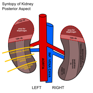
Syntopy of Kidney
The kidneys are responsible for the removal of excess water, salts and wastes of protein metabolism from blood and at the same time retaining nutrients and chemicals to the blood.
Structure[edit | edit source]
They are bean-shaped, convex lateral margin, concave medial margin. Their color is reddish-brown and has measurements about: 10cm length, 5cm width, and 3cm thickness. The renal hilum (portal renalis) lies in the medial margin, a space where blood vessels, nerves and the renal pelvis (and its adipose tissue) enter and leave.
The kidneys are contained into the renal capsule; a tough capsule made of collagenous fibers and connected on the kidney by areolar tissue. As it proceeds medially towards the hilum, it connects with the connective tissue of the vessels which are entering a central recess, the renal sinus.
They have superior and inferior poles. On the superior poles, there are located the suprarenal glands. They are also enclosed in a capsule and in a cushion of pararenal fat. They are separated from the kidneys by a weak fascial septum, thus the 2 structures are not actually attached to each other.
Syntopy[edit | edit source]
The right kidney is lower than the left by 1 rib difference. This is because of the prominence of the inferior side of the liver on the right side of the body. The right kidney is separated from the liver by the hepatorenal recess. Perirenal fascia (or renal fascia envelope), encloses the kidneys and the suprarenal glands and contain the perirenal fat as cushioning for these structures. The anterior part is called prerenal fascia and the posterior retro-renal fascia. Pararenal fat is located behind the retrorenal fascia and the fascia of quadratus lumborum.
Links[edit | edit source]
Related articles[edit | edit source]
Bibliography[edit | edit source]
- MOORE, Keith L – DALLEY, Arthur F. Clinically Oriented Anatomy. 5. edition. Lippincott Williams & Wilkins, 2005. ISBN 0781736390.


