The Major Histocompatibility Complex, HLA
The Major Histocompatibility Complex, or so called MHC, is a molecule present on the surface of the cell membrane encoded by a gene families found in all vertebrates. These molecules put the cells, that they are bound on, into a constant surveillance by the always vigilant Immune System. Studies on the MHC are important for histocompatibility processes during transplantation and for understanding of the Immune System's response against invaders. Without the MHC the Immune System is totally impaired, exposing the organism to any invasion. In humans this Major Histocompatibility Complex refers to the Human Leukocyte Antigen, or so called HLA encoded by genes found on the short (p) arms of the 6th Chromosome. The HLA is the most polymorphic site in the human genome and expression of HLA genes occurs from both alleles for the same locus. The MHC function is to hold on its head domain a fraction of a protein which will be subsequently put under surveillance by lymphocytes and being recognized as self or non self. The protein fractions can occur either from self proteins of a human cell or even by non self proteins taken up by foreign cells that have been phagocytosed and lysed.
The MHC gene family is divided into three subgroups:
- Class I,
- Class II, and
- Class III.
Class I[edit | edit source]
In the case of HLA, these genes encode for protein complexes found on the surface of the plasma membrane on every human nucleated cell. This antigen undergoes recognition process by the Immune System and are categorized as self antigens with no action taken against them. This is called Immunotolerance where the human lymphocytes are programmed not to destruct self antigens so that autoimmunity is prevented. Examples of HLA genes are the HLA-A, HLA-B, HLA-C which encode for the MHC Class I proteins.
- MHC Class I proteins: These proteins contain α (alpha) chains and β2 (beta2) microglobulin with the latter encoded not by HLA genes but by genes located on 15th Chromosome. There are α1, α2, and α3 chains encoded by the genes with the latter being the transmembrane subunit as it is through which the whole MHC molecule is attached on the plasma membrane. As the α chains are complexed with the β2 microglobulin, a self protein is cleaved by cytosolic proteases into fragments that eventually bind on the head domain of the MHC molecule and serve as epitopes. Once the MHC molecule is formed, the peptide epitope is transported and bound on it by the TAP molecule, so called Transporter associated with Antigen Processing. The whole complex is eventually transported from the endoplasmic reticulum on the surface of the cells.
- MHC Class I recognition: As soon as the α3 transmembrane subunit is bound on the membrane then recognition process can commence. The glycoprotein receptor TCD8 of the cytotoxic T lymphocytes, bind on the head domain of the MHC complex (i.e. on the α1 and α2 subunits) and on the epitope. If the epitope is recognized as non self then the receptor is activated, designated as TCD8+, with subsequent undertaking of actions against the destruction of the whole cell intruder.
Class II[edit | edit source]
In the case of HLA, these genes encode for protein complexes found not on the surface of all cells but only on Antigen Presenting Cells, the so called APCs. APCs are called the cells that uptake a foreign particle or cell by endocytosis with their subsequent lysis and consequent formation of foreign protein fragmental epitopes that are eventually introduced on the plasma surface of the APCs. APCs are phagocytes like macrophages, dendritic cells, B lymphocytes and some epithilial lining cells. Class II genes are: HLA-DPA1, HLA-DPB1, HLA-DQA1, HLA-DQB1, HLA-DRA, and HLA-DRB1 which encode for the MHC Class II proteins.
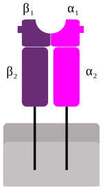
- MHC Class II proteins: The HLA complexes are composed of 4 subunits: 2α and 2β chains all encoded by HLA genes. The head domain is composed of the α1 and β1 subunits that encircle the epitope, whereas the α2 and β2 subunits are both transmembrane with the ability of binding on the cell surface. As soon as the MHC protein is formed, the epitope is ready to be bound on the head domain. In the Class II case, the epitope does not derive from an intracellular protein but from a protein taken up by a non self cell after phagocytosis through lysosomic activity. The MHC Class II molecules involve stimulation of the humoral immunity by stimulating destruction of the invaders but in a rather indirect way in comparison with the Class I.
- MHC Class I recognition: As soon as a macrophage recognizes an MHC Class I complex epitope of an invader cell as a non self, it engulfs the foreign cell and breaks it down in a lytic way through phagocytosis. A protein fragment of this cell is formed as an epitope, which is subsequently placed on a newly synthesized MHC Class II complex as an epitope. The whole molecule is then transferred on the surface on the APC macrophage where the epitope can undergo surveillance by the T helper lymphocytes, designated as Th cell. The glycoprotein receptor of the T helper cell, designated as TCD4 binds on the head domain of the MHC Class II complex (i.e. the α1 and β1 subunits) and on the epitope. When the receptor recognizes the epitope as a non self it is activated and designated as TCD4+. Then the Th cell detaches from the macrophage and attaches on the B lymphocytes inducing their activation, a process that includes proliferation and differentiation of the B cells into plasma cells. The plasma cells will synthesize Immunoglobulins specified for that particular antigenic epitope giving rise to the Adaptive Immunity.
Class III[edit | edit source]
Class III genes are not involved with the Major Histocompatibility Complex although they are located on the 6th Chromosome too. These genes are found between the Class I and II genes and they encode for other immune proteins irrelevant with antigen presentation processing such as TNF-α cytokine.
| Features | MHC Class I pathway | MHC Class II pathway |
|---|---|---|
| MHC - peptide/epitope composition | Polymorphic α chain (α1, α2 and α3), β2 microglobulin, peptide bound to α1 and α2 subunits | Polymorphic chains α (α1 and α2) and β (β1 and β2), peptide binds to both (α1 and β1) |
| MHC cells | All nucleated cells | Macrophages, Dendritic cells, B lymphocytes, some epithelial cells |
| Antigen protein origin | Cytosolic proteins | Proteins inside lysosomes |
| Peptide/epitope generation | Cytosolic proteases | Lysosome acid hydrolases |
| MHC - peptide/epitope binding site | Rough endoplasmic reticulum | Vesicular compartment |
| Immune cell activation | Cytotoxic T lymphocytes, TCD8+ receptor | T Helper cells, TCD4+ receptor |


