Thymus (slide)
Overview[edit | edit source]
Slide 1[edit | edit source]
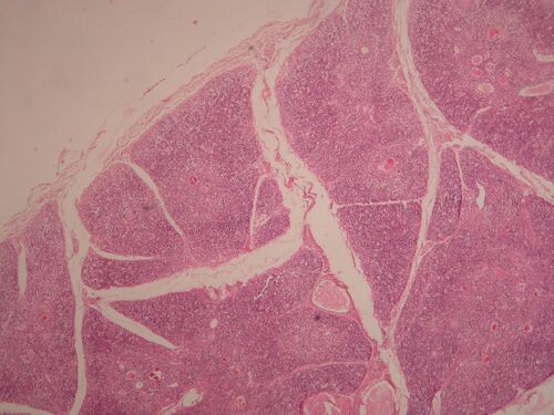
Name: Thymus, lower magnification
Description: The fibrous covering of the thymus penetrates into the interior of the parenchyma and thus creates individual lobules, but they are not completely separated. Each lobule consists of a darker cortex (densely packed T-lymphocytes in a network of reticular epithelial cells) and a lighter medulla (fewer lymphocytes and more reticular epithelial cells). After puberty, the lobules disappear and in the end only residues surrounded by fatty tissue remain.
Slide 2[edit | edit source]
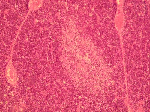
Name: Thymus, part of a lobe
Description: Corresponds to the description in another photo.
Slide 3[edit | edit source]
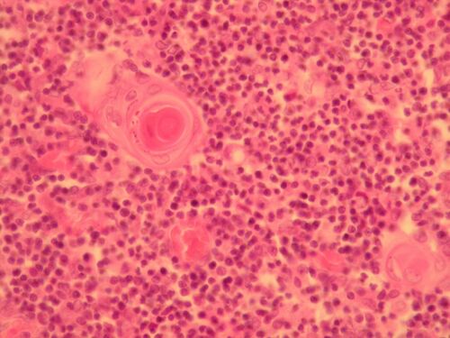
Name: Thymus – pith detail
Description: The thymus does not contain lymph follicles, but its lobules are divided into cortex and medulla only. Here we see in detail the medulla of the thymus, where the specific Hassal bodies (large formation in the upper left quarter) are located, which are composed of concentric layers of reticular epithelial cells. Blood vessels are also found here.
Lymphatic tissue[edit | edit source]
Links[edit | edit source]
Module Cellular Foundations of Medicine (3. LF)


