Trypanosomes
From WikiLectures
| Trypanosoma gambiense | |
| Kinetoplasty (whips) | |
| Trypanosomatidae | |
| Occurrence | West Africa |
|---|---|
| Disease | sleeping sickness , chronic form |
| Infectious
stage and method of infection |
metacyclic trypomastigote - inoculative
(when stabbed with a gloss ) |
| Diagnostics | microscopy |
| Therapy | suramine, arsenic compounds |
| MeSH ID | D014347 |
Trypanosomes are parasitic protozoa belonging to the class of flagella.
Trypanosoma gambiense and Trypanosoma rhodesiense[edit | edit source]
The equivalent name of Trypanosoma gambiense is Trypanosoma brucei
- occurrence : they are connected by the same continent - Africa - but each occurs in a different part:
- T. gambiense: West Africa
- T. rhodesiense: East Africa
- they have a single flagellum that forms an undulating membrane along the body
- they are extracellular parasites
- we find them in blood , lymph and cerebrospinal fluid
- carrier : tse-tse fly or glossina
- source of infection :
- T. gambiense: sick person
- T. rhodesiense: reservoir animals (humans enter the trypanosome life cycle randomly)
![]() In human disease, both species cannot be distinguished
In human disease, both species cannot be distinguished
Life cycle[edit | edit source]
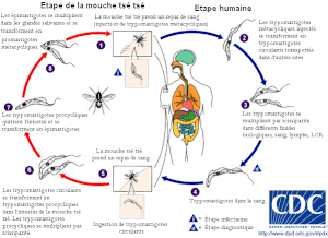
- trypanosomes develop in the gut, sucker and salivary glands of the carrier
- glossina sucks trypomastigotes
- procyclic trypomastigotes travel to the salivary glands and transform into epimastigotes
- at the end of their development, glossins appear in the saliva as so-called metacyclic trypomastigotes = the only stage capable of infecting humans
- as soon as the glosin stings, it releases trypanosomes into human skin = inoculatory transmission
- The sting of a fly is very painful
Disease[edit | edit source]
You can find more detailed information on the Sleeping Disease page.
Trypanosoma cruzi[edit | edit source]
| Trypanosoma cruzi | |
| Kinetoplasty (whips) | |
| Trypanosomatidae | |
| Occurrence | Central and South America |
|---|---|
| Disease | Chagas disease
(American trypanosomiasis) |
| Infectious
stage and method of infection |
metacyclic trypomastigote -
contaminating (from faeces of sucking bedbugs) |
| Diagnostics | microscopy, serology, xenodiagnosis
(sucking of bugs) |
| Therapy | does not exist |
| MeSH ID | D014349 |
- slender whipworm with a wavy body
- invasive, extacellular and intracellular parasite = enters nuclear cells (endothelial cells, muscle cells of all types and neuroglia)
- transmission :
- bugs - especially
- transplacentally
- transfusion (blood)
- orally - rarely - this can happen with insufficient heat treatment of the nine-banded armadillo, from which the bug is infected
Life cycle[edit | edit source]
- Trypanosomes multiply in the intestine of bugs (subfamily Triatominae) in the form of epimastigotes , which turn into infectious trypomastigotes in the rectum of bugs .
- Then the bugs attach to humans → they start sucking (especially at night).
- When sucking bugs , they harden - trypanosomes are present in the feces - the so-called contaminating mode of transmission .
- Trypanosomes are able to actively penetrate the skin .
- Metacyclic trypomastigotes penetrate the body cells of the host, where they multiply intensively as small amastigotes .
- After several divisions, shortly before the cell ruptures, amastigotes turn into trypomastigts, which are released into the bloodstream and initiate infection of other cells.
- Additional blood bugs become infected when the blood is sucked.
Disease[edit | edit source]
You can find more detailed information on the Chagas disease page .
Links[edit | edit source]
External links[edit | edit source]
References[edit | edit source]
- BEDNÁŘ, M, et al. Lékařská mikrobiologie. 1. vydání. Marvil, s. r. o., 1996. ISBN 80-238-0297-6.
- RNDr. Eva Nohýnková, Ph.D. [přenáška z parazitologie]
- VOLF, Petr a Petr HORÁK. Paraziti a jejich biologie. 1. vydání. Praha : Triton, 2007. 318 s. s. 78,80. ISBN 978-80-7387-008-9.


