White matter of the cerebral hemisphere


Contains Info about:
White matter of Cerebral Hemisphere; association and commissural fibers; internal capsule (draw scheme of tracts in internal capsule)
White matter, composed of myelinated fibers, serves as a crucial component within the brain's architecture. Let's delve into its intricate structures:
1. Centrum Semiovale:
- Present in each hemisphere
2. Corpus Callosum:
- This vital structure connects both centrum semiovale regions, facilitating communication between the brain hemispheres.
There are 3 types of white matter fibers:
a) Association Fibers
b) Commisural fibers
c) Projecting Fibers
Association Fibers[edit | edit source]
- These fibers link cortical areas within the same hemisphere, fostering communication between adjacent gyri. - Short Association Fibers: Connecting nearby gyri.
- Long Association Fibers:

- Uncinate Fasciculus: Links the inferior frontal lobe, crucial for motor speech, to the temporal lobe.
- Cingulum: Characterized by long curves, it connects the frontal and parietal lobes with parahippocampal and adjacent temporal cortical regions.
- Superior Longitudinal Fasciculus: - Among the largest, it connects the frontal lobe to the occipital and temporal lobes.
- Inferior Longitudinal Fasciculus: situated deep within the cerebral hemisphere's lateral border. Facilitates communication from the occipital lobe to the temporal lobe, subsequently connecting to the fronto-occipital fasciculus.
- Fronto-Occipital Fasciculus: Extending from the frontal to the occipital lobes.
- Perpendicular fasciculus: A bundle of association fibers running vertically and interconnecting regions of the temporal, occipital, and parietal lobes.
Understanding these structures provides insights into the brain's intricate connectivity, essential for various cognitive functions.
Commissural Fibers[edit | edit source]
Facilitate communication between corresponding regions of the two cerebral hemispheres. The primary structures involved are:
- Corpus Callosum: Serving as the largest commissural structure, it connects both cerebral hemispheres and comprises three parts:
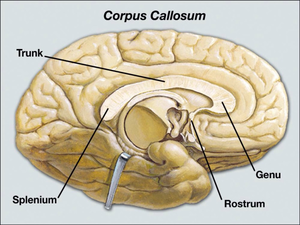 Corpus Collosum[1]
- Rostrum (Anterior)
- Body: The central portion; Curved anterior part forming the forceps minor.
- Splenium (Posterior): The posterior part.
- Anterior Commissure: This structure has two parts:
- Anterior Part: Contains fibers decussating from the olfactory tract, facilitating the sense of smell.
- Posterior Part: Connects the temporal lobes.
- Posterior Commissure: It crosses over the opening of the cerebral aqueduct into the third ventricle. Notably, it plays a crucial role in the pupillary light reflex, where fibers from the pretectal nucleus cross en route to the Edinger-Westphal nucleus.
- Fornix: This structure connects the hippocampus to the mammillary body.
- Habenular Commissure: Crossing superior to the pineal stalk, it connects both habenular nuclei in the epithalamus.

Projecting Fibers[edit | edit source]


Form a compact band known as the internal capsule, consisting of two limbs, anterior and posterior, which continue at the genu.
After emerging superiorly from the internal capsule, these fibers form the corona radiata.
Notably, nerve fibers from the posterior limb radiate to the calcarine sulcus, forming the optic radiation essential for visual processing.
Internal Capsule[edit | edit source]
Descends from Central Semiovale to Diencephalon.
Divides straiatum to caudate and putamen.
References
