Sideropenic anemia: Difference between revisions
(Created page with "__BEZOBSAHU__{{Infobox - onemocnění| česky = Sideropenická anémie| obrázek = Iron deficiency anemia.jpg| popisek = Siderocyty v krevním nátěru | rizikové faktory = m...") |
(Creation of page and migration of article from czech. Initial translation and rewording) |
||
| Line 1: | Line 1: | ||
Sideropenic anemia (also known as Iron-deficiency Anemia) is a condition caused by a disorder of hemoglobin synthesis due to iron deficiency. This is the most common type of [[anemia]] in children.<ref name="KlinPed2012">{{Citace| typ = kniha| příjmení1 = Lebl| jméno1 = J| příjmení2 = Janda| jméno2 = J| příjmení3 = Pohunek| jméno3 = P| kolektiv = ano| titul = Klinická pediatrie| vydání = 1| vydavatel = Galén| rok = 2012| strany = 538-540| rozsah = 698| isbn = 978-80-7262-772-1}}</ref> | |||
= | =Causes of emergence= | ||
''' | '''Insufficient supply''' of [[iron]] in food is rare in the czech republic country. Substances that inhibit resorption (phosphates, phytates) can cause reduced intake. It is more often manifested in malabsorption ([[Celiac disease]], [[Crohn's disease]], after a resection of the stomach or intestines). It also occurs with '''excessive blood loss''' during chronic bleeding ([[peptic ulcers]], [[hemorrhoids]], [[GIT]] cancer, [[menorrhagia]], [[metrorrhagia]]). Iron deficiency also manifests itself if the organism has increased demands for hematopoiesis (during pregnancy, in childhood). | ||
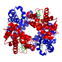
[[File:1GZX Haemoglobin.png|thumb|right|200px| | [[File:1GZX Haemoglobin.png|thumb|right|200px|Hemoglobin structure – Subunits <span style="color:red;">'''α'''</span> and <span style="color:blue;">'''β'''</span> in red and blue, heme containing iron is in green]] | ||
; | ;Causes of sideropenic anemia in children | ||
:''' | :'''Malabsorption''' - treatment with antacids, drugs that increase gastric pH, drinking tea and coffee. | ||
:''' | :'''Insufficient/unavailable iron stores''' - bleeding, [[epistaxis]], gastric and duodenal ulcers, [[Meckel's diverticulum]], [[cow's milk protein allergy]], parasites, Helicobacter pylori infection, esophageal varices, tumors and polyps of the digestive system, inflammatory diseases of the GIT, AV malformations, diverticulitis, hemorrhoids, heavy menstrual bleeding, infections/tumors of the uropoietic tract, pulmonary hemosiderosis | ||
:''' | :'''Congenital/acquired disorders of iron metabolism''' – [[atransferrinemia]], [[disorders of heme synthesis]].<ref name="KlinPed2012" /> | ||
== | ==Iron in the human body== | ||
There are a total of '''3-4 g of iron''' in the body, most of which is found in hemoglobin. Iron recycling from decomposing erythrocytes yields 20 mg per day. In adults, <5% of the total iron comes from the diet, while in an infant it is around 30%. | |||
A full-term newborn has '''sufficient iron reserves for the first 6 months''', after which a dietary supply of iron is necessary. Premature babies have a lower hemoglobin concentration and lower iron reserves, therefore iron depletion already occurs around the 2nd month. Breast milk and cow's milk have the same iron content, but cow's milk is more difficult to absorb. The iron content in breast milk decreases after 5 months of breastfeeding. Boys need a higher supply of iron during puberty due to the increase in muscle mass and myoglobin. Girls need a higher iron intake due to losses during menstrual bleeding.<ref name="KlinPed2012" /> | |||
[[File:Heme_b.svg|thumb|200px|Hemoglobin]] | |||
[[File:Heme_b.svg|thumb | |||
== | ==Clinical picture and diagnosis== | ||
''' | '''Microcytic hypochromic anemia'''. The erythrocyte volume distribution width (RDW) is usually increased due to anisocytosis. Thrombocytosis (500-700×109/l) is relatively common.<ref name="KlinPed2012" /> | ||
#''' | #'''Prelatent sideropenia''' – deficiency of stored iron (reduced [[ferritin]] level, other values in the norm). | ||
#''' | #'''Latent sideropenia''' – deficiency of stored and stored iron in macrophages. This leads to iron deficiency in erythropoiesis (decreased ferritin, iron and increased soluble transferrin receptor sTFR), without anemia. | ||
#'''Sideropenická anémie''' – deficit zásobního i erytrocytárního železa má za následek pokles hladiny hemoglobinu, hematokritu, snížený střední objem erytrocytu (MCV), snížený obsah hemoglobinu v erytrocytu (MCH). Dále se projeví nízkou hladinou železa, ferritinu a zvýšením celkové vazebné kapacity stimulací produkce transferinu v játrech a zvýšení sTFR. Saturace transferrinu klesá pod 16 %, klesá hladina [[hepcidin]]u.<ref name="KlinPed2012" /> | #'''Sideropenická anémie''' – deficit zásobního i erytrocytárního železa má za následek pokles hladiny hemoglobinu, hematokritu, snížený střední objem erytrocytu (MCV), snížený obsah hemoglobinu v erytrocytu (MCH). Dále se projeví nízkou hladinou železa, ferritinu a zvýšením celkové vazebné kapacity stimulací produkce transferinu v játrech a zvýšení sTFR. Saturace transferrinu klesá pod 16 %, klesá hladina [[hepcidin]]u.<ref name="KlinPed2012" /> | ||
There is a '''reduced amount''' of [[reticulocytes]] in the blood, as '''more erythroblasts''' are found in the bone marrow. The marrow is normocellular, and will be hypercellular in anemia due to increased losses. '''Depletion of stored iron''' is characteristic ([[macrophages]] do not show hemosiderosis). It is mainly manifested by tissue [[hypoxia]] (eg '''tiger heart'''). | |||
In children, it manifests itself most often between 6 months and 3 years of age and then in puberty. Iron deficiency during the child's growth period can lead to growth retardation, psychomotor development and cognitive function disorders (negative effect on learning to concentrate). Damage to the epithelium leads to atrophy of the papillae of the tongue and increased brittleness of the nails.<ref name="KlinPed2012" /> | |||
== | ==Differential Diagnosis== | ||
*[[ | *[[Anemia of inflammatory diseases]] – classically normocytic normochromic, in a third of patients microcytic hypochromic, but elevated ferritin and hepcidin. | ||
*[[ | *[[Thalassemia]] - microcytic hypochromic anemia, lower RDW, in the smear typical target-shaped erythrocytes and basophilic stippling of erythrocytes. | ||
*[[ | *[[Lead poisoning]] - microcytic/normocytic normochromic anemia; behavioral changes, convulsions, kidney damage, abdominal pain and vomiting. | ||
*[[ | *[[Sideroblastic anemia]] - hypochromic erythrocytes, increased serum iron, increased transferrin saturation, coronal sideroblasts in the bone marrow (the "coronas" are iron-packed mitochondria).<ref name="KlinPed2012" /> | ||
== | ==Therapy and prevention== | ||
Prevencí je přidávání železa do kojenecké výživy, zavádění masozeleninových příkrmů, cereálie a mléčné přípravky obohacené o železo, vyloučení neadaptovaného kravského mléka z kojenecké stravy a profylaktické podávání železa u nedonošených dětí. | Prevencí je přidávání železa do kojenecké výživy, zavádění masozeleninových příkrmů, cereálie a mléčné přípravky obohacené o železo, vyloučení neadaptovaného kravského mléka z kojenecké stravy a profylaktické podávání železa u nedonošených dětí. | ||
{{Dobrý příklad|Substituce: perorální preparáty železa (nalačno) za současného užívání '''kyseliny askorbové''', která zvyšuje resorpci železa.}} | {{Dobrý příklad|Substituce: perorální preparáty železa (nalačno) za současného užívání '''kyseliny askorbové''', která zvyšuje resorpci železa.}} | ||
| Line 59: | Line 52: | ||
<mediaplayer width="500" height="300">https://www.youtube.com/watch?v=WvD4p8FkQpY</mediaplayer> | <mediaplayer width="500" height="300">https://www.youtube.com/watch?v=WvD4p8FkQpY</mediaplayer> | ||
<noinclude> | <noinclude> | ||
==Odkazy== | ==Odkazy== | ||
===Související články=== | ===Související články=== | ||
Revision as of 22:14, 12 November 2022
Sideropenic anemia (also known as Iron-deficiency Anemia) is a condition caused by a disorder of hemoglobin synthesis due to iron deficiency. This is the most common type of anemia in children.[1]
Causes of emergence
Insufficient supply of iron in food is rare in the czech republic country. Substances that inhibit resorption (phosphates, phytates) can cause reduced intake. It is more often manifested in malabsorption (Celiac disease, Crohn's disease, after a resection of the stomach or intestines). It also occurs with excessive blood loss during chronic bleeding (peptic ulcers, hemorrhoids, GIT cancer, menorrhagia, metrorrhagia). Iron deficiency also manifests itself if the organism has increased demands for hematopoiesis (during pregnancy, in childhood).
- Causes of sideropenic anemia in children
- Malabsorption - treatment with antacids, drugs that increase gastric pH, drinking tea and coffee.
- Insufficient/unavailable iron stores - bleeding, epistaxis, gastric and duodenal ulcers, Meckel's diverticulum, cow's milk protein allergy, parasites, Helicobacter pylori infection, esophageal varices, tumors and polyps of the digestive system, inflammatory diseases of the GIT, AV malformations, diverticulitis, hemorrhoids, heavy menstrual bleeding, infections/tumors of the uropoietic tract, pulmonary hemosiderosis
- Congenital/acquired disorders of iron metabolism – atransferrinemia, disorders of heme synthesis.[1]
Iron in the human body
There are a total of 3-4 g of iron in the body, most of which is found in hemoglobin. Iron recycling from decomposing erythrocytes yields 20 mg per day. In adults, <5% of the total iron comes from the diet, while in an infant it is around 30%.
A full-term newborn has sufficient iron reserves for the first 6 months, after which a dietary supply of iron is necessary. Premature babies have a lower hemoglobin concentration and lower iron reserves, therefore iron depletion already occurs around the 2nd month. Breast milk and cow's milk have the same iron content, but cow's milk is more difficult to absorb. The iron content in breast milk decreases after 5 months of breastfeeding. Boys need a higher supply of iron during puberty due to the increase in muscle mass and myoglobin. Girls need a higher iron intake due to losses during menstrual bleeding.[1]
Clinical picture and diagnosis
Microcytic hypochromic anemia. The erythrocyte volume distribution width (RDW) is usually increased due to anisocytosis. Thrombocytosis (500-700×109/l) is relatively common.[1]
- Prelatent sideropenia – deficiency of stored iron (reduced ferritin level, other values in the norm).
- Latent sideropenia – deficiency of stored and stored iron in macrophages. This leads to iron deficiency in erythropoiesis (decreased ferritin, iron and increased soluble transferrin receptor sTFR), without anemia.
- Sideropenická anémie – deficit zásobního i erytrocytárního železa má za následek pokles hladiny hemoglobinu, hematokritu, snížený střední objem erytrocytu (MCV), snížený obsah hemoglobinu v erytrocytu (MCH). Dále se projeví nízkou hladinou železa, ferritinu a zvýšením celkové vazebné kapacity stimulací produkce transferinu v játrech a zvýšení sTFR. Saturace transferrinu klesá pod 16 %, klesá hladina hepcidinu.[1]
There is a reduced amount of reticulocytes in the blood, as more erythroblasts are found in the bone marrow. The marrow is normocellular, and will be hypercellular in anemia due to increased losses. Depletion of stored iron is characteristic (macrophages do not show hemosiderosis). It is mainly manifested by tissue hypoxia (eg tiger heart).
In children, it manifests itself most often between 6 months and 3 years of age and then in puberty. Iron deficiency during the child's growth period can lead to growth retardation, psychomotor development and cognitive function disorders (negative effect on learning to concentrate). Damage to the epithelium leads to atrophy of the papillae of the tongue and increased brittleness of the nails.[1]
Differential Diagnosis
- Anemia of inflammatory diseases – classically normocytic normochromic, in a third of patients microcytic hypochromic, but elevated ferritin and hepcidin.
- Thalassemia - microcytic hypochromic anemia, lower RDW, in the smear typical target-shaped erythrocytes and basophilic stippling of erythrocytes.
- Lead poisoning - microcytic/normocytic normochromic anemia; behavioral changes, convulsions, kidney damage, abdominal pain and vomiting.
- Sideroblastic anemia - hypochromic erythrocytes, increased serum iron, increased transferrin saturation, coronal sideroblasts in the bone marrow (the "coronas" are iron-packed mitochondria).[1]
Therapy and prevention
Prevencí je přidávání železa do kojenecké výživy, zavádění masozeleninových příkrmů, cereálie a mléčné přípravky obohacené o železo, vyloučení neadaptovaného kravského mléka z kojenecké stravy a profylaktické podávání železa u nedonošených dětí.
- Substituční přípravky
- Soli dvojmocného železa – síran železnatý (Aktiferrin, Ferronat);
- železo v komplexu s polysacharidy (Maltofer) – méně nežádoucích účinků;
- parenterální podávání trojmocného Fe – při prokázané malabsorpci, značných krevních ztrátách, noncompliance atd.. K úpravě anémie dochází stejně rychle jako při perorální substituci.
- Nežádoucí účinky
- Pocit plnosti, nauzea, zácpa či průjem. Akutní otrava železem se projeví závažnými gastrointestinálními příznaky, systémovou toxicitou. Jako antidotum používáme Template:HVLP, který váže toxické volné železo.
Efekt léčby se hodnotí vzestupem retikulocytů (4. – 10. den léčby), postupným vzestupem hemoglobinu (o 20 g/L za 4 týdny). Pro doplnění zásob železa je obvykle nutné pokračovat celkem 3–5 měsíců.[1]
Souhrnné video
<mediaplayer width="500" height="300">https://www.youtube.com/watch?v=WvD4p8FkQpY</mediaplayer>
Odkazy
Související články
Reference
Použitá literatura
- ČEŠKA, Richard – ŠTULC, Tomáš. Interna. 2. edition. 2015. 909 pp. ISBN 978-80-7387-895-5.
- PASTOR, Jan. Langenbeck's medical web page [online]. [cit. 12.4.2010]. <http://langenbeck.webs.com>.


