Ventricular fibrillation: Difference between revisions


| (9 intermediate revisions by 3 users not shown) | |||
| Line 1: | Line 1: | ||
__noTOC__ | __noTOC__ | ||
[[File:Heart of ventricular fibrillation.png|thumb|Scheme of chaotically arising excitations in the myocardium of the heart chambers during ventricular fibrillation.]] | [[File:Heart of ventricular fibrillation.png|thumb|Scheme of chaotically arising excitations in the myocardium of the heart chambers during ventricular fibrillation.]] | ||
Ventricular fibrillation (VF) and rapid ventricular tachycardia (VT) belong to the so-called malignant [[arrhythmias]]. These are [[heart rhythm disorders]] that urgently threaten the patient's life (they must be dealt with immediately) and are the most common cause of [[Sudden cardiac death|sudden cardiac death]] (NSS).<ref name="Mlčochová ">{{ | Ventricular fibrillation (VF) and rapid ventricular tachycardia (VT) belong to the so-called malignant [[arrhythmias]]. These are [[heart rhythm disorders]] that urgently threaten the patient's life (they must be dealt with immediately) and are the most common cause of [[Sudden cardiac death|sudden cardiac death]] (NSS).<ref name="Mlčochová ">{{Cite | ||
| type = article | | type = article | ||
| surname1 = Mlčochová | | surname1 = Mlčochová | ||
| name1 = Hank | | name1 = Hank | ||
| | | others = yes | ||
| article = Catheter ablation of ventricular fibrillation using elimination of triggering ventricular ectopy | | article = Catheter ablation of ventricular fibrillation using elimination of triggering ventricular ectopy | ||
| journal = Interv Akut Kardiol | | journal = Interv Akut Kardiol | ||
| url = http://www.solen.cz/pdfs/kar/2007/05/02.pdf | | url = http://www.solen.cz/pdfs/kar/2007/05/02.pdf | ||
| year = 2007 | | year = 2007 | ||
| | | the_year = 5 | ||
| volume = 5 | | volume = 5 | ||
| pages = 173-176 | | pages = 173-176 | ||
| Line 16: | Line 16: | ||
}}</ref> Ventricular fibrillation occurs relatively often as part of an acute myocardial infarction. | }}</ref> Ventricular fibrillation occurs relatively often as part of an acute myocardial infarction. | ||
== Etiology == | == Etiology == | ||
The causes of ventricular fibrillation include:<ref name="Mlčochová"/> <ref name="Haman">{{ | The causes of ventricular fibrillation include:<ref name="Mlčochová"/> <ref name="Haman">{{Cite | ||
| type = web | | type = web | ||
| | | surname1 = Haman | ||
| name1 = Peter | | name1 = Peter | ||
| url = http://www.ekg.kvalitne.cz/tvorba.htm#Fibrillation%20komor | | url = http://www.ekg.kvalitne.cz/tvorba.htm#Fibrillation%20komor | ||
| | | source_name = EKG Learning Site | ||
| subtitle = ECG Basics | | subtitle = ECG Basics | ||
| cited = 2010-11-21 | | cited = 2010-11-21 | ||
| Line 30: | Line 30: | ||
== Pathogenesis == | == Pathogenesis == | ||
In the case of ventricular fibrillation, the individual [[Myocardium (histological preparation, HE)|cardiomyocytes]] of the heart chambers contract completely '''unsynchronized'''. These chaotic and rapid contractions lead to a complete '''failure of the heart's function as a pump''. Since the heart is not pumping any blood ([[Determining cardiac output|minute cardiac output]] is almost zero), there is severe '''perfusion failure'''' of vital organs including the [[brain|cerebrum]] (→ loss of [[Consciousness and his disturbance|consciousness]] occurs within 10 s) and without immediate [[Basic emergency resuscitation|cardiopulmonary resuscitation]] and [[defibrillation]], ventricular fibrillation leads to death (irreversible brain damage develops in 3-5 minutes<ref>{ { | In the case of ventricular fibrillation, the individual [[Myocardium (histological preparation, HE)|cardiomyocytes]] of the heart chambers contract completely '''unsynchronized'''. These chaotic and rapid contractions lead to a complete '''failure of the heart's function as a pump''. Since the heart is not pumping any blood ([[Determining cardiac output|minute cardiac output]] is almost zero), there is severe '''perfusion failure'''' of vital organs including the [[brain|cerebrum]] (→ loss of [[Consciousness and his disturbance|consciousness]] occurs within 10 s) and without immediate [[Basic emergency resuscitation|cardiopulmonary resuscitation]] and [[defibrillation]], ventricular fibrillation leads to death (irreversible brain damage develops in 3-5 minutes<ref>{{Cite | ||
| type = book | | type = book | ||
| surname1 = Czech | | surname1 = Czech | ||
| name1 = Richard | | name1 = Richard | ||
| | | others = yes | ||
| title = Intern | | title = Intern | ||
| | | edition = 1 | ||
| | | location = Prague | ||
| publisher = Triton | | publisher = Triton | ||
| year = 2010 | | year = 2010 | ||
| Line 46: | Line 46: | ||
[[File:Ventricular fibrillation video.webm|preview|video in English: definition, pathogenesis, symptoms and complications, diagnosis, treatment]] | [[File:Ventricular fibrillation video.webm|preview|video in English: definition, pathogenesis, symptoms and complications, diagnosis, treatment]] | ||
<big>'''Ventricular fibrillation:'''</big> {{Video | url=https://youtu.be/z3cnSQmJxvc?t=1225}} | |||
<big>'''Ventricular flutter:'''</big> {{Video | url=https://youtu.be/1hEbgdYVaxI?t=1059}} | |||
<big>'''Ventricular flutter 2:'''</big> {{Video | url=https://youtu.be/z3cnSQmJxvc?t=1131}} | |||
Physiologically, a depolarization wave occurs in the [[Cardiac conduction system|SA node]]. In ventricular fibrillation, '''abnormal electrical impulses'' occur directly in the heart's chambers. These abnormal impulses can arise unifocally or multifocally, it can be a reentry mechanism or a combination of these mechanisms. | Physiologically, a depolarization wave occurs in the [[Cardiac conduction system|SA node]]. In ventricular fibrillation, '''abnormal electrical impulses'' occur directly in the heart's chambers. These abnormal impulses can arise unifocally or multifocally, it can be a reentry mechanism or a combination of these mechanisms. | ||
| Line 85: | Line 92: | ||
* [[Cardiac conduction system]] | * [[Cardiac conduction system]] | ||
=== External links === | === External links === | ||
* [https://www. | * [https://www.ecgbook.com/ventricular-fibrillation/ Ventricular fibrillation and ECG] - Free ECG book | ||
=== References === | === References === | ||
| Line 94: | Line 102: | ||
* ČESKA, Richard, et al. Internal 1st edition. Prague: Triton, 2010. 855 pp. pp. 480-481. <nowiki>ISBN 978-80-7387-423-0</nowiki>. | * ČESKA, Richard, et al. Internal 1st edition. Prague: Triton, 2010. 855 pp. pp. 480-481. <nowiki>ISBN 978-80-7387-423-0</nowiki>. | ||
=== | === References === | ||
* | * HAMPTON, John R. ''EKG Concise, Clear, Clear. ''6th edition. Grada, 2005. 149 pp. ISBN 80-247-0960-0 . | ||
* HAMAN, Peter. ''EKG Learning Website : EKG Basics'' [online]. [feeling. 2010-11-19]. < http://www.ekg.kvalitne.cz/tvorba.htm#Fibrillace%20komor >. | |||
* HELLO, Robert. Cardiology ring. III. internal clinic of VFN and 1st Faculty of Medicine of the University of Prague in Prague, 2009. | |||
* VILIKUS, Zdeněk. Interpretation of ECG at rest and during exercise. Institute of Physical Education Medicine 1. LF UK and VFN; 2010. | |||
* | |||
* | |||
* VILIKUS, Zdeněk. | |||
[[ | [[Category:Cardiology]] | ||
[[ | [[Category:Physiology]] | ||
[[ | [[Category:Pathophysiology]] | ||
[[ | [[Category:Internal medicine]] | ||
Latest revision as of 10:38, 25 September 2024
Ventricular fibrillation (VF) and rapid ventricular tachycardia (VT) belong to the so-called malignant arrhythmias. These are heart rhythm disorders that urgently threaten the patient's life (they must be dealt with immediately) and are the most common cause of sudden cardiac death (NSS).[1] Ventricular fibrillation occurs relatively often as part of an acute myocardial infarction.
Etiology[edit | edit source]
The causes of ventricular fibrillation include:[1] [2]
- Organic heart disease (CHD and MI, cardiomyopathy, cardiac amyloidosis, ... ),
- long QT syndrome (LQTS), Brugado syndrome,
- idiopathic ventricular fibrillation.
Pathogenesis[edit | edit source]
In the case of ventricular fibrillation, the individual cardiomyocytes of the heart chambers contract completely unsynchronized'. These chaotic and rapid contractions lead to a complete failure of the heart's function as a pump. Since the heart is not pumping any blood (minute cardiac output is almost zero), there is severe perfusion failure' of vital organs including the cerebrum (→ loss of consciousness occurs within 10 s) and without immediate cardiopulmonary resuscitation and defibrillation, ventricular fibrillation leads to death (irreversible brain damage develops in 3-5 minutes[3]).
Ventricular fibrillation:
Ventricular flutter:
Ventricular flutter 2:
Physiologically, a depolarization wave occurs in the SA node. In ventricular fibrillation, 'abnormal electrical impulses occur directly in the heart's chambers. These abnormal impulses can arise unifocally or multifocally, it can be a reentry mechanism or a combination of these mechanisms.
Ventricular tachycardia, ventricular flutter or early ventricular extrasystole can become ventricular fibrillation.
Clinical manifestation[edit | edit source]
Clinically, ventricular fibrillation manifests as:[2]
- loss of consciousness;
- inaudible heart sounds;
- non-palpable pulse;
- immeasurable pressure.
Diagnostics[edit | edit source]

The diagnosis is based on the ECG.
Individual cardiomyocytes contract completely independently and unsynchronized, therefore we cannot identify any QRS complexes on the ECG. We see irregular waves with a frequency of 300/min and more.
Treatment[edit | edit source]
- Treatment consists of immediate initiation of cardiopulmonary resuscitation'' (opinions on precordial beat vary) and external or internal defibrillation as soon as possible.
- Some types of ventricular fibrillation can be treated with radiofrequency ablation' of focal ventricular ectopy.[1]
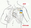
- In primary and secondary prevention, some antiarrhythmic drugs and implantation of an implantable cardioverter-defibrillator (ICD) can be used.

Portable manual external defibrillator

Implantable cardioverter-defibrillator
Scheme of the established ICD

X-ray image of an implanted ICD


Links[edit | edit source]
Related Articles[edit | edit source]
External links[edit | edit source]
- Ventricular fibrillation and ECG - Free ECG book
References[edit | edit source]
- MLČOCHOVA, Hanka, et al. Catheter ablation of ventricular fibrillation by elimination of triggering ventricular ectopy. Interv Akut Kardiol [online] . 2007, year 5, vol. 5, pp. 173-176, also available from <http://www.solen.cz/pdfs/kar/2007/05/02.pdf>. ISSN 1803-5302.
- HAMAN, Peter. EKG Learning Website : EKG Basics [online]. [feeling. 2010-11-21]. < http://www.ekg.kvalitne.cz/tvorba.htm#Fibrillation%20komor >.
- ČESKA, Richard, et al. Internal 1st edition. Prague: Triton, 2010. 855 pp. pp. 480-481. ISBN 978-80-7387-423-0.
References[edit | edit source]
- HAMPTON, John R. EKG Concise, Clear, Clear. 6th edition. Grada, 2005. 149 pp. ISBN 80-247-0960-0 .
- HAMAN, Peter. EKG Learning Website : EKG Basics [online]. [feeling. 2010-11-19]. < http://www.ekg.kvalitne.cz/tvorba.htm#Fibrillace%20komor >.
- HELLO, Robert. Cardiology ring. III. internal clinic of VFN and 1st Faculty of Medicine of the University of Prague in Prague, 2009.
- VILIKUS, Zdeněk. Interpretation of ECG at rest and during exercise. Institute of Physical Education Medicine 1. LF UK and VFN; 2010.
- ↑ Jump up to: a b c MLČOCHOVÁ, Hank. , et al. Catheter ablation of ventricular fibrillation using elimination of triggering ventricular ectopy. Interv Akut Kardiol [online]. 2007, y. 5, vol. 5, p. 173-176, Available from <http://www.solen.cz/pdfs/kar/2007/05/02.pdf>. ISSN 1803-5302.
- ↑ Jump up to: a b HAMAN, Peter. EKG Learning Site : ECG Basics [online]. [cit. 2010-11-21]. <http://www.ekg.kvalitne.cz/tvorba.htm#Fibrillation%20komor>.
- ↑ CZECH, Richard, et al. Intern. 1. edition. Prague : Triton, 2010. 855 pp. pp. 480-481. ISBN 978-80-7387-423-0.
