Bones of the upper limb


In general, the limb bones ( ossa membrorum ) are divided into four parts:
- cingulum (braid), by which the limb is attached to the axial skeleton;
- stylopodium – proximal part formed by one bone ( humerus = arm bone, femur );
- zeugopodium – the distal part formed by two bones (on the upper limb it is the radius and ulna , on the lower tibia and fibula );
- autopodium – arm and leg skeleton.
Overview of bones[edit | edit source]
The bones of the upper limb ( ossa membri superioris ) form:
Armband[edit | edit source]
The braid of the upper limb consists of:
The clavicle is articulated with the sternum , forming the only skeletal connection with the axial skeleton.
Arm[edit | edit source]
The arm ( brachium ) is formed by a single bone, the humerus , which is articulated with the girdle.
Forearm[edit | edit source]
The forearm ( antebrachium ) consists of two bones:
These two bones are very closely related. Rotational movements of the radius around the ulna allow pronation and supination of the hand.
Hand[edit | edit source]
The hand ( manus ) is made up of several groups of small bones:
- carpal bones – (os scaphoid, os lunate, os triquetrum, os pisiforme, os trapezium, os trapezoide, os capitate, os hamatum)
- metacarpal bones – (five metacarpal bones, numbered 1 to 5 from radial to ulnar side)
- finger bones – (each finger has three bones, the thumb has only two)
Scapula[edit | edit source]
__

The scapula (shoulder bone, shoulder blade, wing bone) is a flat bone, triangular in shape. It is placed on a posterolateral aspect of the thoracic cage, ranging from the level of the second rib to the level of the seventh rib.
Scapula connects with the humerus (upper arm bone) in the glenohumeral (shoulder) joint and the clavicula (collar bone) in the acromioclavicular joint. There is no direct connection between the thoracic cage and scapula. It is held in place thanks to the surrounding muscles.
The name scapula comes from early Roman times, meaning a small shovel.
Functions[edit | edit source]
The scapula is the origin and instertion of various muscles
The scapula protects the thoracic cavity form the dorsal side
Moving the upper limb[edit | edit source]
As is, the humerus can only move 90° upwards, into the horizontal plane. Moving any more, the humerus' head would collide with the coracoid process of the scapula. To move the upper limb above the horizontal plane, the coracoacromial ligament (fornix humeri) needs to be engaged.
The scapula and humerus move in a 1:2 ratio. When the upper limb is abducted 180°, 60° of those 180 occur by rotation of the scapula and 120° by movement of the humerus itself.
Borders[edit | edit source]
The three borders of the scapula are:
- Margo medialis
- Medial border, parallel to the longitudinal axis of the spine
- Margo lateralis
- Lateral, or axillary border
- Margo superior
- Superior border
Angles[edit | edit source]
Borders of the scapula connect in three angles:
- Angulus superior
- Superior and medial border
- Angulus inferior
- Medial and lateral border
- Angulus lateralis
- Lateral and superior border
Surfaces[edit | edit source]
Dorsal surface[edit | edit source]
The back of the scapula is divided into two unequal parts by the spine of the scapula . The spine ends in a process called the acromion. The acromion forms the cavity of the glenohumeral joint.
The portion of the dorsal surface above the spine is called the supraspinous fossa. M. supraspinatus originates there.
The portion of the dorsal surface below the spine is called the infraspinous fossa. It is much larger than the supraspionous fossa. M. infraspinatus originates there.
Both muscles are parts of the rotator cuff, rotating the humerus and providing stability for the glenohumeral joint.
The coracoid process originates from the superior border.
|
|---|

Ventral surface[edit | edit source]
The front of the scapula has a concavity called the subscapular fossa. M. subscapularis originates in the fossa.
|
|---|

Lateral surface[edit | edit source]

The glenoid cavity can be found at the lateral angle. The acromion and the coracoid process make its top border.
Above and below the cavity, two tubercles serve as origins of muscles:
- Supraglenoid tubercle
- Origin of the long head of m. biceps brachii
- Infraglenoid tubercle
- Origin of the long head of m. triceps brachii
References[edit | edit source]
Scapula. Scapula [online]. [cit. 2019-11-09]. Dostupné z: https://www.wikiskripta.eu/w/Scapula
Scapula. In: Wikipedia: the free encyclopedia [online]. San Francisco (CA): Wikimedia Foundation, 2019 [cit. 2019-11-09]. Dostupné z: https://en.wikipedia.org/wiki/Scapula
HUDÁK, Radovan a David KACHLÍK. Memorix anatomie. 4. vydání. Praha: Triton, 2017. ISBN 978-80-7553-420-0.
ČIHÁK, Radomír. Anatomie. Třetí, upravené a doplněné vydání. Praha: Grada, 2016. ISBN 978-80-247-3817-8.
Ossification[edit | edit source]
Ossification of the scapula begins at about the 8th week of pregnancy, gradually other ossification centers are formed in the processus coracoideus , acromion and fossa glenoidalis . From the 14th to the 20th year, individual ossification centers coalesce.
clavicula[edit | edit source]
The clavicle is a long bone , but it is only about 15 cm long. Its body is curved like an ace (lateral third dorsally, the rest ventrally).
- The medial end , extremitas sternalis , is larger and articulates with the manubrium of the sternum .
- The lateral end , extremitas acromialis , is articulated with the upper arm of the scapula .
The ligamentum coracoclaviculare attaches to the tuberositas coracoidea at the lower end of the clavicle . The clavicle is the only bony connection between the axial skeleton and the upper limb. All pressures and impacts from the upper limb to the chest are transmitted through it. Fractures of the clavicle tend to be much more common than disruption of one of the two joints involved.

Ossification[edit | edit source]
The clavicle begins to ossify around the 6th week of pregnancy. The peculiarity is that the bone ossifies endesmally . Thanks to this, ossification is completed more quickly, the body of the clavicle is already bony in a newborn. Complete completion of ossification occurs around 21 years of age.
Humerus[edit | edit source]
The arm bone ( humerus ) measures about 30 cm, it is a typical long bone. It can be divided into:
- caput − head,
- corpus − body of the bone,
- condylus − distal part.

Caput[edit | edit source]
The caput humeri is covered by cartilage on the medial part and forms the head of the shoulder joint . Just below the edges of the cartilage is the collum anatomicum (anatomical neck). There are two bumps on the front under the head:
- tuberculum majus − laterally,
- tuberculum minus − ventrally.
Between the tubercles is the sulcus intertubercularis , and the tendon of the long head of the biceps passes through it . Below the level of both bumps is the site of the most common fractures of the humerus, the collum chirurgicum . The head of the humerus is turned dorsally by 30° relative to the axis of the condyles, which is in the frontal plane (retroversion).
Corpus[edit | edit source]
The triangular body of the humerus connects to the head. The tuberositas deltoidea roughness is visible at the point of attachment of the deltoideus muscle . The sulcus nervi radialis can be seen at the place where the radial nerve runs (the nerve can be injured in the case of a humerus fracture in this place). In the middle of the length of the body there is a foramen nutricium , through which nourishing vessels enter the bone.
Condyle[edit | edit source]
It forms the most distal part of the humerus. The articular surfaces located on it participate in the elbow joint . The ovoid condyles extend into:
- epicondylus lateralis,
- epicondylus medialis - behind it is the sulcus nervi ulnaris (for nervus ulnaris , popularly known as "bruise"; in case of fractures of the condyles, this nerve can therefore be injured).
Just above the articular surfaces are three depressions:
- fossa radialis − ventrally, laterally;
- fossa coronoidea − ventrally, medially, fits into it at the bend of the processus coronoideus of the ulna ;
- fossa olecrani − on the dorsal side, the olecranon fits into it .
On the most distal part there are two articular surfaces:
- capitulum humeri − head, laterally, articulated with the radius ;
- trochlea humeri − pulley, medially, articulated with the ulna .
Ossification[edit | edit source]
From the 3rd week of pregnancy, ossification begins in the diaphysis and both epiphyses of the humerus. Proximal growth cartilage is more active than distal. Smaller ossification nuclei are also formed (for example in bumps). Around the age of 20, the last growth spurts disappear.
Ulna[edit | edit source]
The elbow bone ( ulna ) is located on the medial side of the forearm ( ulnar direction ). On the proximal part, there is an articular surface, forming a fossa of the medial ulnar and proximal radioulnar joints. The body of the bone ( corpus ulnae ) has a triangular cross-section. The distal end of the ulna ( caput ulnae ) is indirectly articulated with the carpus and directly with the radius .
Anatomy[edit | edit source]
Proximal part[edit | edit source]
- It creates a notch, incisura trochlearis , in the anatomical position it is open ventrally, into which the trochlea humeri fits , and is bordered by the following formations:
- olecranon – the most proximal part of the bone, when the joint is flexed it forms the tip of the elbow;
- processus coronoideus – distal border of the notch;
- incisura radialis – a small depression covered by cartilage, connected to the articular surface, into which the caput radii fits .
The body of the ulna[edit | edit source]
- The body has a triangular cross-section , the edges are reversed ventrally, dorsally and laterally ( margo anterior, posterior et interosseus ).
- Margo interosseus faces the radius and is connected to it by an interosseous ligamentous membrane.
- The foramen nutricum is directed proximally.
The head of the ulna[edit | edit source]
- The head of the ulna is formed by the cylindrical articular surface of the circumferential articularis , which laterally attaches to the radius and proximally to the discus articularis .
- From the mediodorsal side, the processus styloideus ulnae emerges , which is easily palpable.
Clinical contexts[edit | edit source]
See Forearm Fractures for more detailed information .


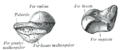
Radius[edit | edit source]
The spindle bone ( radius ) is located on the lateral side of the forearm (radial direction). It is similar in shape to the ulna , but narrower proximally than distally. Aiming for the thumb.
We divide it into three parts:
- caput radii – the proximal part of the cylindrical shape;
- corpus radii – triangular body;
- distal part – the distal end of the radius.

Radius – anterior view

Radius – posterior view
Caput radii[edit | edit source]
- The head of the spindle bone has a barrel shape and is entirely covered by articular cartilage ;
- the proximal surface is articulated with the capitulum humeri ;
- the annular articular surface of the circumference of the radii fits into the notch on the ulna;
- the head of the radius is connected to the corpus radii by the neck ( collum radii ).
Corpus radii[edit | edit source]
- The body of the spindle bone has three edges ( margo anterior, posterior et interosseus );
- the interosseous edge is connected to the edge of the same name on the ulna by a fibrous membrane, the membrana interossea antebrachii ;
- on the body there is a tuberosity ( tuberositas radii ) where the biceps brachii muscle attaches ;
- the foramen nutricium is located about halfway up the bone and is directed proximally.
Distal part of radius[edit | edit source]
- It is a wider surface ( facies articularis carpalis radii ) covered by articular cartilage that participates in the radiocarpal articulation ;
- the distal part of the ulna fits into a small notch here ;
- the distal part of the radius rotates about the axis formed by the ulna during pronation and supination .
Ossa carpi[edit | edit source]
The carpal bones ( ossa carpi ) form two rows of small bones, each row of four:
- proximal row – (mediolateral, ulnoradial) os pisiforme, triquetrum, lunate, scaphoid ;
- distal row – (mediolateral, ulnoradial) os hamatum, capitatum, trapezoideum, trapezium .
The proximal row participates in the radioulnar articulation. The ossa metacarpi (metacarpal bones) mount on the distal one .
The carpal bones form the bony base of the carpal tunnel , through which the tendons of the flexos digitorum superficialis et profundus and the median nerve pass , which is often compressed here ( carpal tunnel syndrome ).
Proximal row[edit | edit source]
the pisiform[edit | edit source]
Pea bone. It has the shape and size of a pea. It develops as a sesamoid bone in the wrist flexor ulnar tendon .
The mouth of a trident[edit | edit source]
Triangular bone. It has the shape of an irregular tetrahedron.
A crescent moon[edit | edit source]
Calendula bone. Its shape resembles a crescent moon.
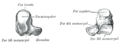
Os scaphoideum[edit | edit source]
Navicular bone (not to be confused with the os naviculare of the tarsus ). Its shape remotely resembles a boat, the depression is turned medially . It is the largest bone of the proximal row. It bears the tuberculum ossis scaphoidei . (unfortunately, the English version of the picture contains the error just mentioned - naviculare x scaphoideum)
Distal row[edit | edit source]
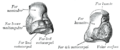
Hooked mouth[edit | edit source]
Hook bone. It has a triangular shape. The hamulus ossis hamati (hook) protrudes prominently from it .
Capped mouth[edit | edit source]
Skull bone. The largest carpal bone. The proximally facing part is called the caput ossis capitati and fits into the proximal row between the os lunate and the os scaphoideum .
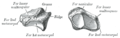
Trapezoidal bone/Polygonal bone less[edit | edit source]
A small polygonal, or scarf-shaped, bone. Its shape resembles the letter L ( shoe ), in the sagittal cross-section it has the shape of a pyramid turned with its base on the dorsum of the manus .
Trapezoidal face/Major polygonal face[edit | edit source]
Large polygonal bone. It has a similar shape to the trapezoid axis , but is larger. Laterodorsally, there is a conspicuous saddle-shaped surface for articulation with the first (thumb) metacarpal bone.
Ossification[edit | edit source]
The carpal bones do not ossify until postnatally in this order (approximate time of ossification in parentheses ):
- os capitatum (2. m), hooked (3. m), triquetrum (3. r), lunate (4. r), scaphoid (5. r), trapezium (5. r), trapezoid (6. r), pisiform (7.-13. r) .
Ossification of the carpal bones is a good indicator of a child's physical maturity.
Pictures[edit | edit source]

Left pisiform bone

The left triquetrum

Left crescent moon
Left scaphoid bone
Left hook mouth
Left-headed mouth

Left trapezoidal bone
Left trapezius bone

The relative position of the bones

The bones of the hand


Ossa metacarpi[edit | edit source]


The skeleton of the palm (metacarpus) is made up of five bones (ossa metacarpi). These correspond in shape to long bones, but are classified among short bones (around 10 cm). Metacarpals are located between the carpal bones (carpus) and the finger joints (phalanges), where they are articularly connected to both the carpal bones and the finger joints. All joints are strengthened by ligaments on both sides. Each of the bones then has its characteristic feature, for example, metacarpal axis I is the shortest. We distinguish three parts:
- basis – proximal expanded end with articular socket, articulates with carpus;
- corpus - body (corresponds to the diaphysis);
- caput – distal extension of the bone, the ossa digitorum is attached to it.
All bones are well palpable from the dorsal side. They are numbered from I. to V. in the radioulnar direction.
Finger bones[edit | edit source]
The skeleton of the fingers consists of the ossa digitorum (manus) or phalanges , the joints of the fingers (hands), which are two on the thumb and three on the other fingers. There are three main sections on each article:
- basis phalangis , the base of the joint – wider proximal section,
- corpus phalangis , the body of the joint – the middle slender part,
- caput phalangis , the head in which the joint ends distally.
The bases of the joints are transversely expanded, on the proximal side there is a concave articular surface for the adjacent bone (see below). The bodies of the finger joints are dorsally slightly convex, palmarly straight to slightly concave. The heads are the convex surfaces of the pulley joints with a corresponding groove; the relevant guide bar is based on the following article.
- The articles differ according to the position of the finger:
- The phalanx proximalis (prima) is the longest, the bases of the proximal joints have a pit proximally, transversely oval, for the head of the metacarpal .
- The medial phalanx (secunda) is shorter than the first phalanx, on the edges it has fine edges for the attachment of the flexor tendons. The thumb has no phalanx media.
- Phalanx distalis (tertia) is the shortest; on the palm side near the base it has a roughened place for the tendon of the flexor digitorum longus; distally, it ends with an extension - the tuberositas phalangis distalis is a roughness on the palm side of the final extension of the distal joints, and a dense ligament is attached to it , which fills the distal end of the belly of the finger.
All articles are palpable , especially from the dorsal side. Only the base can be felt from the distal articles.
Ossa sesamoidea manus – sesame bones of the hand[edit | edit source]
Ossa sesamoidea manus are small bones at the metacarpophalangeal joints. Two are constantly found, on either side of the metacarpophalangeal joint of the thumb , and not infrequently they may also be present at other metacarpophalangeal joints . They are formed in the tendons of the muscles that tighten in those places. At the second to fifth metacarpophalangeal joints, they are often only cartilaginous (without ossification ), and therefore escape attention during an X-ray examination. The os pisiforme carpus is also considered to be a bone of sesame origin.
links[edit | edit source]
[edit | edit source]
References[edit | edit source]
- CIHÁK, Radomír. Anatomy. 2nd edition. Prague: Grada Publishing, as, 2008. p. 516. ISBN 80-7169-970-5 .
Bones bones of the skull bones of the neurocranium os occipitale • os sphenoidale • os ethmoidale • os temporale • os frontale • os parietale • os lacrimale • os nasale • vomer bones of the splanchnocranium maxilla • os palatinum • os zygomaticum • mandible • os hyoideum • ossicula auditus • concha nasalis inferior axial skeleton spine • vertebrae • ribs • sternum • os sacrum bones of the upper limb plait scapula • clavicle arm and forearm humerus • ulna • radius hand carpus • metacarpus • finger bones bones of the lower limb plait os coxae ( hip bone • ischium • pubic bone ) thigh and lower leg femur • patella • tibia • fibula leg ossa tarsi • ossa metatarsi • bones of the fingers Portal: Anatomy
