Chromosomal Abnormalities
(Redirected from Chromosome abnormalities)
Chromosomal abnormalities – sometimes also called cytogenetic disorders – are very common. Although we don´t see many affected people. How it this possible? Most fetus with some chromosomal abnormality usually do not survive. About 50% of first–trimester abortions is connected with some cytogenetic mistake. The incidence of chromosomal abnormalities is approximately 1 out of 200 of newborns.[1].
We recognize two types of chromosomal abnormalities:
- numeric;
- structural.
We are able to find the disorders due to karyotype testing. The cytogeneticists get the samples (blood, amnionic fluid), then stain the chromosomes and arrange them in the right order by their length. Then we can see some changes in pattern (banding). The most typical method is Giemsa stain, so it is usually called "G banding". This is important for structural abnormalities. Numeral abnormalities we can identify even easier.
Other classification of chromosomal abnormalities depends on fact which type of chromosome is affected – autosomes (Down syndrome, DiGeorge syndrome) or sex chromosomes (Klinefelter syndrome, Turner syndrome).
Types of Structural Abnormalities
Translocation
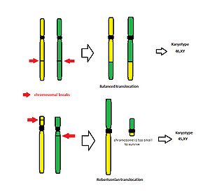
- During translocation, a part of one chromosome is transferred to another chromosome.
- It is very important whether the translocation is balanced or unbalanced. Balanced means that two chromosomes just exchange their parts but the number of chromosomes(46 chromosomes) as well as no loss of genetic material stays the same.
- Fortunately – typical place of break is near the centromere, usually only small arms of the acrocentric chromosomes are lost. There are no crucially important genes coded by these chromosomal segments. So, a carrier of such Robertsonian translocation can normally survive this cytogenetic change.
- Unfortunately problems occur during the fertilization with a gamete of a carrier of a balanced translocation. Carrier of the translocation may produce unbalanced zygotes, because the process of homologous chromosomes pairing during meiosis is interrupted. This is very important because unbalanced gametes lead to abnormalities in offspring. The reason is that the offspring receives altered chromosome from the carrier which may lack several important genes.
- Therefore the only clinical symptom found in the carriers of balanced translocations may be the reproduction failure.
Types of translocation
- reciprocal – translocation between two chromosomes ("A segment" goes to "B chromosome" and "B segment" goes to "A chromosome")
- Robertsonian – translocation (or fusion) of two acrocentric chromosomes
Deletion
- Deletion is characterized by the loss of a part of chromosome. Two breaks have to occur for deletion of the interstitial segment. For deletion of terminal segment (telomere) one break is enough. Segments which were deleted from the chromosome are not able to "live" on their own and the genes present in those segments are lost.
- One special example of deletion exists. It is called "ring chromosome". It is a situation when chromosome lost both of its ends. The long and the small arms then connect together and chromosome became a ring shaped.
Isochromosomes
- Isochromosomes are created by the incorrect division of centromere. Normally centromere divides vertically. In this case it divides horizontally.
- The result is usually the loss of one arm. It means that newly created chromosome has just two long arms or two short arms which are normally connected by centromere.
- It occurs relatively frequently in X chromosome.
- It is a huge problem during the fertilization. Because fetus then becomes trisomic for one arm and monosomic for the second arm.
Inversion
- For inversion are typical two breakages in the different part of the chromosome. The newly created segments then replace each other.
- Inversion was discovered in 1921.
- Although we still don´t know why inversion exists, we know that it is the most important mechanism of reorganizing of the genome.
- We recognize 2 types of inversion:
- pericentric – causing deletions, insertions or abnormal centromeres;
- paracentric – more common type, it is less harmful for its carrier.
- Inversion suppresses the recombination process.
How We Denote Parts of a Chromosome
When we want to describe the location of some structural abnormality we used special marks. We have to count the regions always from the centromere – for both arms. Example: 1q13
- the first number is a number of chromosome – chromosome 1;
- (p) and (q) are used for marking of arm – (p) for small arm and (q) for long arm – the long arm;
- the third number is for the region of chromosome – region 1;
- last number is for the band – band 3.
So the change is located on the long arm of chromosome 1 (see Fig. Chromosomal marking).
Etiology of Chromosome Abnormalities
Etiology of chromosome abnormalities is pretty variable. The most often reason is mistake which occurs during the cell division. It is connected with wrong development of the sperm or ovum (female reproduction cell). We recognize two types of cell division – mitosis and meiosis. Another causes are the maternal age and the ifluence of the enviromental.
Chromosome abnormalities are usually fatal. Each second first-trimester abortion is caused by them. Children who survive and get born suffer from very serious mental and physical problems. The screening for chromosome abnormalities is very important. The cytogeneticists use the karyotype testing.
Problems in the Cell Division
We recognize two main types of the cell divison. The first one is the meiosis. It is the process of division of reproduction cells. The result is a cell with 23 chromosomes (it is haploid). Fetus get 23 chromosomes from the mother and 23 chromosomes from the father. Cells of fetus are already diploid (2N). Mistake during meiosis leads to incorrect number of the chromosomes in the egg or in the sperm. Child can get some extra chromosome (trisomy) or miss it (monosomy).
The second type of cell division is the mitosis. It occurs in all non-reproductive cells. It is a form of duplication of the genetic information, followed by the halving of material. The parent cell has 92 chromosomes (4N), two subsidiary cells have 46 chromosome each (2N). Mitosis starts immediately after fertilization and continues throught whole life. When mistake occurs, the chromosomes may not be equal. Problems in mitosis leads to the mosaicism more often.
Age of Parents
We are sure about the strong influence of the maternal age (especially in Down syndrome). The paternal age is less important, but still has its importance. The difference is in cell division of reproduction cells. The number of eggs (female´s cells) is done by our birth. So eggs underway meiosis many times. The later age of delivery means the higher risk of some abnormalities. It takes just 72 hours to the developement of sperm cells. It is less propably to make a mistake during this period.
To the older women is recommended to visit some genetic counseling centre. "The older women" means more than 35 years. The prenatal diagnosis is the best way to find some affected children. The method of prenatal diagnosis as an amniocentesis.
Influence of the Environment
It is very hard to tell how important is the enviromental. We can´t find any significant differences between parents with child with a chromosome abnormality and parents with healthy child. They have usually very similar lifestyle or habits. But there are still some dangerous influences – X-rays, medication or food. Most of them have a cumulative character.
Because we are not sure about the origin of abnormalities, it is hard to recommend any prevention. Sometimes it is said that the folic acid has a positive role in prevention of congenital abnormalities. Pregnant women should also get vitamines to reduce risks.
Links
Related articles
- Karyotype
- Disorders of the Autosomes
- Disorders of the Sex Chromosomes
- The Sex Chromosomes and Sex Determination
Sources
- KUMAR, ABBAS, FAUSTO, MITCHELL,, et al. Robbins Basic Pathology. 8th edition edition. 2007. ISBN 978-0-8089-2366-4.
References
- ↑ About 50% of first–trimester abortions is connected with some cytogenetic mistake. The incidence of chromosomal abnormalities is approximately 1 out of 200 of newborns


