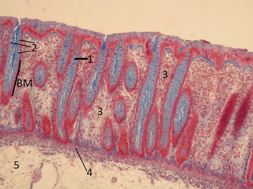
Colon (SFLT)
Colon (AZAN)[edit | edit source]
Description: The epithelium of the large intestine is single-layered and cylindrical . With AZAN staining, nuclei are stained red and mucin in goblet cells blue. Epithelial cells in the lamina propria (sparse collagenous tissue) form simple tubular glands (crypts of Lieberkühn).
Colon (HE)[edit | edit source]
Description: The mucosa of the large intestine on an oblique section. Cross-sections of Lieberkühn's crypts are visible. The lighter cells are goblet cells. Mucin is not colored during normal processing. The spaces between the crypts are filled by sparse collagenous tissue with a large number of free cells (lymphocytes, plasma cells, etc.)
Colon (PAS)[edit | edit source]
Description: The PAS reaction stains polysaccharides, glycoproteins and proteoglycans pink. In the specimen, the mucin in the goblet cells of the epithelium and, to a lesser extent, the intercellular mass in the ligament are stained by the PAS reaction.
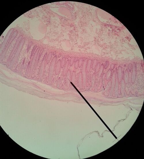
Colon (HE)[edit | edit source]
Description: Large intestine - small magnification. In addition to the lamina mucosa (lamina epithelialis - epithelium; lamina propria mucosae - sparse collagenous tissue; lamina muscularis mucosae - smooth muscle), the lamina submucosa' is also visible here, also formed by sparse tissue, which, however, is much denser compared to the lamina propria, i.e. it contains more fibers and fewer cells.
Colon, lamina submucosa - plexus submucosus[edit | edit source]
Description: The submucosus plexus (a small ganglion in the center of the visual field) is embedded in a thin collagenous ligament. It contains multipolar neurons surrounded by satellite cells and connected by nerve fibers.
Colon (PAS and hematoxylin staining)[edit | edit source]
Description: PAS will show polysaccharides, proteoglycans and glycoproteins in the tissue (cyclam pink). In the preparation, PAS is positive mucin in goblet cells (single cell intraepithelial glands).


