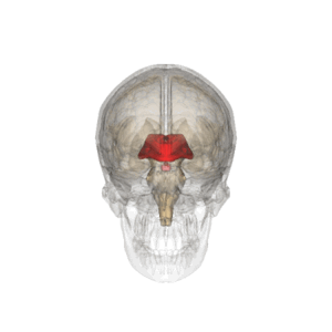
Diencephalon
Diencephalon also called midbrain consists of 5 functionally and morphologically distinct parts. Dorsoventrally they are: epithalamus, thalamus, metathalamus, subthalamus and hypothalamus.
Anatomy[edit | edit source]
The midbrain connects to the upper end of the brain stem. It is located between the hemispheres of the terminal brain, thus it is not well visible. The only visible structure lies on the ventral surface of the brain, and that is the hypothalamus. The posterior border is formed by the upper end of the interpeduncular fossa, or two bumps, the corpora mamillaria. It ends in the area of the optic chiasm.
The diencephalon is formed by further brain development of the anterior cerebral sac (prosencephalon), in which the original division to allar a basal discs is evident. The thalamus (sensitive structure) and basal hypothalamus (visceromotor structure) develop from the alar plates.
Description[edit | edit source]
The most conspicuous part of the midbrain are the two arches, which are the thalami that form the lateral wall of the '''IIIrd ventricle'''. Furthermore the choroidal fibrous bodies of the IIIrd ventricle form the ceiling of the 3rd ventricle. The place of attachement of the choroidal body is called taenia thalami.
The diencephalon contains the 3rd cerebral ventrical, which is the continuations of the aquaeductus mesencephali, coming from the IV.th cerberal ventricle. It then flows into the intraventricular foramina, through which it enters the lateral ventricles (between the hemispheres of the terminal brain).
The medial wall of the diencephalon (side walls of the III. ventricle) is divided by a paired groove - sulcus hypothalamicus (corresponds to sulcus limitans of the neural tube). This structure divides the diencephalon into dorsal and ventral parts. The dorsal part includes the thalamus, metathalamus and epithalamus, which are mainly sensitive (sensory). The ventral part includes the subthalamus and hypothalamus, whose functions are mainly motoric.
Epithalamus[edit | edit source]
Is the dorsocaudal part of the diencephalon, which consists of habenular nuclei and pineal body. The habenular nuclei are contained in the habenular trigone, which is formed by the extension of the bundle of white matter (stria medullaris thalami). Both trigones together form the habenula, within which fibers of the of the stria medullaris thalami cross. In this area of crossing the pineal body (epiphysis) extends from the epithalamus.
- Nuclei
- Inside the habenula there are habenular nculei (nucleus habenularis medialis et lateralis). Their activity is somatomotor and visceromotor, allowing reactions of olfactory and limbic arousal. Habenula is a functional part of the limbic system.
- Tracts
- Commissura posterior connects posterior thalamic nuclei, colliculi superiores and pretectal nuclei of both sides. It contains fibers emerging from the ncl. interstitialis, ncl. Darkshevichi, pretectal nuclei and a part of habenulotektal fibers.
Thalamus[edit | edit source]
Thalamus presents a paired part of the diencephalon and it is oval in shape
The anterior part narrows to the anterior tubercule and posterior rounded part is referred to as the pulvinar. The 2 parts of the thalamus are
joined to each other through the interthalamic adhesion.
Metathalamus[edit | edit source]
The metathalmus occipitally follows the thalamus. It consists of the lateral geniculate body, which is located under the pulvinare and mediale. The metathalamus is involved in the visual pathway and auditory pathway, receiving signals from the mesencephalon.
- Nuclei
- Ncl. corporis geniculati lateralis belongs to the visual pathway and ncl. corporis geniculati medialis belongs to the auditory pathway.
Subthalamus[edit | edit source]
Lies ventrally from the thalamus and laterally from the hypothalamus.
Hypothalamus[edit | edit source]
A small part of the diencephalon is found under the thalamus. Rostrally it reaches up to the lamina terminalis and caudally to the posterior margin of the mamillary bodies. It lies laterally to the III. cerebral venrticle and medially to capsula interna. The infundibulum protrudes on the base of the hypothalamus and continues as a stalk on which the hypophysis (pituitary gland) hangs.
Hypothalamus serves as the highest viscermotoric center in the human body. Further on it is the center of activities of the autonomic nervous system. Its functions include alsoendocrine activities.
Links[edit | edit source]
Related articles[edit | edit source]
Literature used[edit | edit source]
- PETROVICKÝ, Pavel. Anatomie s topografií a klinickými aplikacemi III.. 1. edition. Martin, SR : Vydavateľstvo Osvěta, 2002. vol. III.. Chapter Neuroanatomie, smyslová ústrojí a kůže. ISBN 80-8063-048-8.
- GRIM, Miloš – DRUGA, Rastislav. 4a. Centrální nervový systém. 2. edition. Praha : Galén, 2002. ISBN 978-80-7262-938-1.


