Epiphysis
This article is about the pineal gland. The article Epiphysis (part of the bone) discusses the part of the bone.
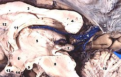
Epiphysis (cerebral appendage, pineal gland, epiphysis cerebri, corpus pineale) is up to 1 cm long and 3–5 mm wide at its widest point, a conical unpaired structure, weighing approx. 120 mg. It forms the largest part of the diencephalic structure called epithalamus . The epiphysis is attached to the commissura habenularum, but extends caudally as far as between the mesencephalic colliculi superiores.[1] Functionally, it is an endocrine gland'.
Development[edit | edit source]
The pineal gland arises as a protrusion from the proliferation of ependymal cells in the posterior part of the ceiling plate of the diencephalon around the 7 week (in the front part, the cells of the ceiling plate participate in the formation of the tela choroidea venticuli tertii). During this period, mesenchyme also grows into the foundations of the future pineal gland. Around the 14 week or later, the empty space in the original foundation is filled and the cells differentiate into characteristic elements.
Construction[edit | edit source]
On the surface of the pineal gland is a weak fibrous capsule connected to the pia mater (capsula corporis pinealis). From it, thin fibrous septa penetrate into the body of the epiphysis, which divide the organ into irregular, incomplete lobes formed by beams. Along the septa, blood vessels and unmyelinated nerve fibers enter the pineal gland. Epiphyseal cells can be divided into two groups:
- own pinealocytes (pinealocyti cardinales),
- interstitial neuroglial cells' of the type astrocytes (gliocytes pineales).
Pinealocytes predominate in number.
Pinealocytes are highly modified neurons arranged in trabeculae with fenestrated capillaries running between them. They are stellate in shape and emit numerous cytoplasmic projections with dilated club-like endings near the vessels. Small electron-dense granules are concentrated in these dilatations. Pinealocytes are producers of melatonin, a derivative of serotonin.
Interstitial glial cells have nuclei that are almost rod-shaped, much more stainable (with a higher content of heterochromatin). When observed with a light microscope, it is sometimes possible to observe their stronger cytoplasmic protrusions.
Numerous 'unmyelinated axons then end between the pinealocytes. Their endings sometimes have the character of special synapses. A large number of vesicles with norepinephrine can be found here, and serotonin has also been demonstrated here.[2]
Calcified, often lamellar concretions, so-called ``brain sand (``acervulus cerebri) are typical for the pineal gland. The concretions have an irregular shape and are made of a substance of unknown protein origin. They turn red when stained with hematoxylin-eosin. Ca2+ salts, mainly hydroxyapatite and calcium carbonate, are deposited soon, mainly on the periphery of often compound concretions. Such concretions then acquire a purple to blue tint.
Changes during life[edit | edit source]
The number of grains of cerebral sand increases with age, so that while during the first ten years of life they can be found in approximately 12% of the pineal glands, in older people it is already 70-80%. Since puberty, the pineal gland undergoes "degenerative changes". Not only more concretions appear, but also more ligaments originating from the septa. The melatonin-producing parenchyma decreases, so melatonin production also decreases with age. In older people, it is only about a 'quarter of the value measured at a young age.
Function[edit | edit source]
The pineal gland is a producer of "melatonin", thereby participating in the regulation of circadian rhythms. Melatonin acts as a specific hormone, but at the same time affects and modulates the function of a number of endocrine glands. It affects the rhythmic function of the gonads and pituitary gland (positively affects the production of growth hormone and growth factors). Melatonin secretion fluctuates over the course of 24 hours, as it is dampened by light. Tumors in the region of the pineal gland are associated with precocious puberty (pubertas praecox) and hypertrophy of the gonads in pediatric patients.
Phylogeny[edit | edit source]
Today's human pineal gland evolved from the 'third parietal eye' of reptiles living millions of years ago. In today's reptiles (in some such as the New Zealand Hateria) it has a photosensitive function, and in amphibians melatonin is a very effective regulator of pigmentation (a lighter skin tone can be achieved by clustering melanin granules in melanocytes). [3]
Links[edit | edit source]
Related Articles[edit | edit source]
External links[edit | edit source]
References[edit | edit source]
- ↑ PETROVICKY, Pavel. Anatomy with Topography and Clinical Applications : III. volume, Neuroanatomy, sensory system and skin. 1. edition. Osveta, 2002. 542 pp. ISBN 80-8063-048-8.
- ↑ KONRÁDOVÁ, Václava – CARBON, George – VAJNER, Luděk. Functional Histology. 2. edition. H & H, 2000. 291 pp. ISBN 80-86022-80-3.
- ↑ Original unpublished script by Prof. R. Jelínek and the team. Script of histology and embryology
References[edit | edit source]
- Original unpublished script by Prof. R. Jelínek and the team. Script of histology and embryology
- SADLER, Thomas, W. Langmanova lékařská embryologie. 1. czech edition. Grada Publishing, a. s, 2011. 414 pp. ISBN 978-80-247-2640-3.
- KONRÁDOVÁ, Václava – UHLÍK, Jiří – VAJNER, Luděk. Functional histology. 2. edition. H & H, 2000. 291 pp. ISBN 80-86022-80-3.
- NOVOTNÁ, Božena – MAREŠ, Jaroslav. Developmental Biology for Physicians. 1. edition. Karolinum, 2005. 99 pp. ISBN 80-246-1023-X.
- PETROVICKÝ, Pavel. Anatomie s topografií a klinickými aplikacemi : III. bundle, Neuroanatomy, sensory system and skin. 1. edition. Osveta, 2002. 542 pp. vol. 3. ISBN 80-8063-048-8.


