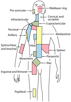
General anatomy of the lymphatic system
The lymphatic system or lymphatic system' consists of lymphatic vessels and highly specialized lymphoid organs and tissues, primarily thymus, spleen (lien) and tonsillae (tonsillae). The smallest lymphatic vessels are called lymphatic capillaries and run along arteries and veins. All lymphatic vessels merge into two large channels, the thoracic lymphatic duct' (ductus thoracicus) and the right lymphatic duct' (ductus lymphaticus dexter), which flow into venae brachiocephalicae near heart. Lymph is thus drained from the tissues by the lymphatic system into blood. There is a sap, which is a product of tissue fluid. In order for the lymph to return to circulation, 2-3 liters of this fluid must pass daily through the lymph capillaries into the bloodstream.
Lymphatic vessels[edit | edit source]
The beginning of lymphatic capillaries is observed blindly in the intercalated connective tissue in almost all organs (except central nervous system, bone, cartilage, teeth and those without blood supply – e.g. epithelium of the skin). Lymphatic capillaries connect with each other anastomoses in a network from which lymphatic collectors are collected. The aquifers already resemble blood vessels (also referred to as vasa lymfatica), while the walls of the lymphatic capillaries are very thin and highly permeable, so that large molecules and particles, including bacteria, that cannot enter the blood capillaries, are carried away by lymph. These collectors usually guide the lymph to the adjacent lymph nodes, which filter the lymph node. From here, the final lymphatic trunks are collected, which lead to larger collectors or veins.
Construction of individual sections[edit | edit source]
The lymphatic capillaries are similar to the blood capillaries with their single layer endothelium, but they are significantly more permeable due to the absence or imperfection of [lamina basalis]]. Thanks to the large slits, the lymphatic capillary is able to accommodate even large molecules impenetrable to capillaries (e.g. chylomikra) and thus provides another transport function of the lymphatic system.
The lymphatic collectors are larger vessels containing characteristic valves arranged in pairs that prevent retrograde lymph flow in physiological cases. The lymphatic collector wall itself already contains tunica intima, media and adventicia, and thus resembles blood vessels.
Lymphatic trunks are already similar to small veins.
Central lymphatic organs[edit | edit source]
The main feature of these organs is their stroma, which forms the reticular epithelium.
Thymus[edit | edit source]
Primary lymphatic organ important especially in childhood. The main function is to acquire immunocompetence and autotolerance T-lymphocytes, which takes place here.
Bone marrow[edit | edit source]
Atypical lymphatic organ ranked among the primary only by some authors. The stroma is reticular connective tissue, which is a sign of rather peripheral lymphatic organs. There is the formation of formed blood elements hemopoiesis.
Peripheral lymphatic organs[edit | edit source]
Lymph[edit | edit source]
These are lymphatic organs of egg-shaped shape of different sizes, forming a network throughout the body. Lymph nodes are mainly located near large arteries and where arteries are palpable below the surface of the body, for example, in the groin, in the armpit and in the neck. They act as sap flow filters and scavenge various foreign particles such as dust, germs or tumor cells.
The entrance to the lymph node is referred to as hilum nodi lymphatici, it is located on the concavity of the node and enters the blood vessels that nourish the organ. In addition to blood vessels, there is a vas efferens – a drainage vessel for lymph from the nodule, which is usually one. On the other hand, the vasa afferentia, which brings the lymph into the nodule and there are more of them, enters the nodule on the convex surface.
On the surface of the nodule we find the connective tissue capsule capsula nodi lymphatici. Below the capsule is the wall itself with beams of connective tissue – trabeculae with numerous macrophages. These beams immerse themselves in the interior of the nodule, dividing the internal tissue into incompletely closed partitions. A network of reticular cells and fibers (reticulum) departs from the beams, which in their spaces form nodal sinuses for lymph flow. These spaces are filled with lymphocytes and thus form the immune centers of the lymph follicles located in the node..
The nodule is divided into three spaces:
- cortex – cortex with lymph nodules, area containing B-lymphocytes, which form the circumference of the nodule;
- paracortical zone – area containing T-lymphocytey, the area between cortex and medulla;
- medulla – medulla, between the paracortical zone and the hilum nodes. Here the medullary sinuses pass into the finite, terminal sinuses, which continue to the vas efferens.
Spleen[edit | edit source]
Organ functional for blood circulation and immunity of the organism.
Diffuse lymphoid tissue in organs[edit | edit source]
Folliculi lymphatici scattered mainly in the digestive tract, upper respiratory tract and urinary tract occur as separately lying folliculi lymphatici solitarii. Folliculi lymphatici aggregati are then grouped nodules in the small intestine. By construction, they resemble lymph nodules in a large nodule. The predominant representatives of immune cells are B-lymphocytes.
The system of these nodules is unstable with the possibility of constantly rebuilding and changing.
Tonsils[edit | edit source]
Lymphatic organs that have highly concentrated lymphoid tissue in the mucosa. They are the primary contact points with antigens in food intake and inhalation of air, therefore they are of great importance shortly after the birth of an individual. In chronic disease, microbes can be infected and serve as their refuge. Therefore, the removal of tonsils can be approached – tonsillectomy. The tonsils are collectively described as the Waldeyer's lymphatic circuit, to which they belong:
- tonsillae palatinae;
- tonsilla lingualis;
- tonsilla pharyngea;
- tonsillae tubariae.
Lymph[edit | edit source]
Lymph is a colourless product of tissue fluid, into which the filtrate of blood capillaries (due to increased filtration and reduced resorption during the capillary) and the fluid with cell metabolites on the other hand passes. It contains a blood plasma composition with a protein content of around 20 g/l. However, this does not apply in the intestinal lymphatic bed, where absorbed fats get into the sap. As a result, the sap present here is whitish to yellowish with a protein content of up to 60 g/l; it is referred to as chylus.
In addition, the lymphatic contains a number of lymphocytes, the amount of which is constantly increasing through the lymphatic vessel system.
Lymph movement is allowed:
- contractions of smooth muscle in medii lymphatic vessels;
- contractions of adjacent skeletal muscles;
- breathing movements;
- peristalsis of the intestine;
- activities of heart.
Functions of the lymphatic system[edit | edit source]
The lymphatic system acts as a drainage system for lymph from tissue fluid and metabolites of cells, which works due to the fact that lymphatic vessels and macromolecular substances, such as chylomikra, can lead from the digestive system due to the permeability of capillaries. The system contains many immune centers, thus significantly contributing to the body's immune defense. ![]() .
.
Links[edit | edit source]
Related Articles[edit | edit source]
- Lymphatic vessels • Lymph node
- Thymus • Spleen
- Head and neck lymphatic system • Lymph nodes of the genitourinary system
- Atlas of histological slides/Lymphatic system
- Lymphatic system on the portal of anatomy
- Lymphatic drainage of limbs
Bibliography[edit | edit source]
- ČIHÁK, Radomír. Anatomie 3. 2. edition. Grada, 2004. 673 pp. pp. 172-178. ISBN 80-247-1132-X.
- TROJAN, Stanislav. Lékařská fyziologie. 4. edition. Grada, 2003. 772 pp. pp. 261. ISBN 80-247-0512-5.
- WESTON, Trevor. Atlas lidského těla. - edition. Levné knihy KMa, 2003. ISBN 80-7321-092-4.
- JUNQUEIRA, L. Carlos – CARNEIRO, Chosé. Základy histologie. 7. edition. H&H, 1999. ISBN 8085787377.
- LÜLLMANN-RAUCH, Renate. Histologie. 1. edition. Grada, 2012. ISBN 978-80-247-3729-4.


