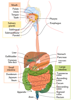
General structure of the alimentary canal
The digestive system (systema digestorium) serves to process nutrients and absorb them . During digestion, food passes through the digestive tract, where it is ground up and chemically broken down into simpler substances. These get into the blood and then into the tissues and cells of the body, where they are used as a source of energy or a building block of cells .
The digestive system consists of a digestive tube and accessory glands - gll. salivariae majores ( glandula parotis , glandula submandibularis , glandula sublingualis , liver and pancreas )
Main organs and sections of the digestive system[edit | edit source]
- cavitas oris - oral cavity
- Dentes - teeth , lingua - tongue , palatum - palate , glandulae oris - salivary glands of the mouth
- Pharynx - pharynx
- Oesophagus - Oesophagus
- Gaster - stomach
- Intestinum tenue - small intestine
- Duodenum - duodenum
- jejunum - the stomach
- ileum - iliac crest
- Intestinum crassum - large intestine
Glands opening into the duodenum
The individual layers of the alimentary canal[edit | edit source]
Tunica mucosa[edit | edit source]
It lines the cavities of the alimentary canal as a soft, pink to red layer, moistened with secretions from the glands. In some sections it is smooth, in others it is folded into cilia or extends into small projections.
- Epithelial lamina
- Formed at the beginning and end of the alimentary canal by a stratified squamous non-keratinizing epithelium in places that serve mainly as mechanical protection (oral cavity, pharynx, esophagus, anal part of the alimentary canal), in the middle parts it is a single-layered cylindrical epithelium, which performs secretory and resorptive function in the intestine.
- It can form eyelashes ( plicae ), papillae ( papillae ) or villi ( villi ).
- Epithelial cells produce mucus and a number of agents that are responsible for digestion, resorption of substances and removal of harmful, indigestible and excess metabolites.
- Lamina propria mucosae
- It is located under the epithelium and is a layer of thin collagenous tissue in which numerous blood and lymphatic vessels and glands are found.
- Lymphatic follicles ( folliculi lymphatici solitarii et aggregati ) are present in some places .
- Just below the epithelium is a zone where there are numerous macrophages , lymphocytes and plasma cells producing antibodies ( immunoglobulins ), which are bound to secretory proteins produced by the epithelial cells and are released onto the mucosal surface.
- Lamina muscularis mucosae
- It is made up of smooth muscle cells.
- It does not always have to be, it allows the mucosa to move along the muscles of the alimentary canal.
Tunica submucosa[edit | edit source]
In other words, the submucosal tissue formed by a thin collagenous tissue in which blood and lymphatic vessels are found . This is the layer connecting the mucous membrane to the muscle. Glands (eg in the duodenum gll. duodenales seu Brunneri or esophagus) and lymphatic tissue also appear in the submucosa . There are also plexuses of stronger blood vessels and nerves (plexus submucosus Meissneri) .
Tunica muscularis externa[edit | edit source]
The muscle layer is formed at the beginning (to the middle of the esophagus) and at the end of the digestive tube by striated muscle , in the middle part, i.e. from the middle part of the esophagus to the lower part of the rectum, by smooth muscle . The muscle is arranged in two layers. The inner layer is circular (stratum circulare) and the outer longitudinal (stratum longitudinale). Both smooth and striated circular muscle is strengthened in places into sphincters, sphincters. Between the circular and longitudinal muscles is a weak layer of ligaments, in which lie other blood and lymphatic vessels and a nerve plexus (plexus myentericus Auerbachi).
Tunica externa[edit | edit source]
The outer surface layer is formed either as a tunica adventitia , composed of thin or thickened tissue, which passes into the surrounding tissue ( mediastinum , paraproctia). The outer layer created in this way covers the parts of the alimentary canal that do not lie in the peritoneal cavity, i.e. cervical and thoracic section of the esophagus and recta . The second form of surface layer is the smooth and shiny tunica serosa , which covers the part of the alimentary canal facing the abdominal cavity. It consists of one layer of flat epithelial cells, the so-called mesothelium cells, underlain by a small layer of subserous tissue, the tunica subserosa.
Links[edit | edit source]
Related resources[edit | edit source]
Practicing histological preparations
References[edit | edit source]
- PASTOR, Jan. Langenbeck's medical web page [online]. [cit. 17.04.2010]. <https://langenbeck.webs.com/>.
- ČIHÁK, Radomír – GRIM, Miloš. Anatomie. 2., uprav. a dopl edition. Praha : Grada Publishing, 2002. 470 pp. vol. 2. ISBN 80-7169-970-5.
- JUNQUIERA, L. Carlos – CARNEIRO, José – KELLEY, Robert O.. Základy histologie. 1. edition. Jinočany : H & H 1997, 1997. 502 pp. ISBN 80-85787-37-7.
- LÜLLMANN-RAUCH, Renate. Histologie. 1. edition. Praha : Grada, 2012. ISBN 978-80-247-3729-4.
- NAŇKA, O., ELIŠKOVÁ M. Přehled anatomie. Druhé, doplněné a přepracované vyd. Praha: Galén - Karolinum, 2009, 416 s. ISBN 978-80-246-1717-6
- ZDN, Trávicí soustava, MUDr. Petr Bednář, 2010 [online] Dostupné na: <https://web.archive.org/web/20160331222721/http://zdravi.e15.cz/clanek/priloha-pacientske-listy/travici-soustava-449015>


