Tongue
Tongue (lingua, glossa) is an organ in the oral cavity helping the mechanical processing of food and serving as an organ of taste and speech.
Anatomical structure of the tongue[edit | edit source]
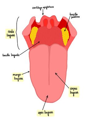
The tongue is a muscular organ covered by a mucous membrane. Using its muscles it is connected to the lower jaw, uvula, soft palate, styloid process of the temporal bone and to the wall of the pharynx. The mucous membrane passes from the lower surface of the tongue into the mucous membrane of the floor of the mouth.
On the tongue we recognize:
- radix linguae (root of the tongue) – the part facing the pharynx;
- corpus linguae (body of the tongue) – hl. part, rests on the palate when the mouth is closed;
- apex linguae (tip of the tongue) – movable front part.
The following are described:
- dorsum linguae (back of the tongue) – then on it:
- sulcus medianus linguae – longitudinal median furrow;
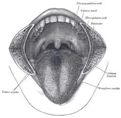
- sulcus terminalis – "V"-shaped groove, separates the corpus from the radix;
- foramen caecum linguae – depression in the middle of the tip of sulcus terminalis, during embryonic development, the base of the thyroid gland separated from here and descended caudally as ductus thyreoglossus;
- margo linguae – lateral edge of the tongue;
- facies inferior – lower surface;
- plica fimbriata – mucosal fold on facies inferior, rest of sublingua;
- frenulum linguae – frenulum of the tongue;
- caruncula sublingualis – in front on both sides of the frenula, the duct of submandibular gland and duct of major sublingual gland opens here;
- plica sublingualis – fold, a large number of duct of minor sublingual gland opens here;
- plica glossoepiglotica mediana – middle, unpaired fold extending from the root of the tongue to the epiglottis;
- plicae glossoepigloticae laterales – right and left paired folds;
- valleculae epiglotticae – 2 pits between the eyelashes.
Mucous membrane of the tongue[edit | edit source]
The mucous membrane of the tongue is covered by a multi-layered squamous epithelium , on the back and tip of the tongue, the epithelium extends in papillae linguales (their epithelium is cornified):
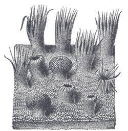
- papillae filiformes (filiform papillae) – the most common, slender, frayed at the end, the epithelium on the surface is subject to keratinization, an important function in grinding the bite when the tongue moves against the hard palate;
- papillae conicae (conical) – sharp, few in humans, common in animals (rough tongue of cats, etc.);
- papillae fungiformes (mushroom like) – club-shaped, individually between papillae filiformes , red (capillaries in the connective tissue show through the thinner epithelium);
- papillae foliatae (leaf-like) – rudimentary, on the margo linguae at the back, in their epithelium we find taste buds;
- papillae vallatae (walled) – the largest, in the walls of the taste buds (caliculi gustatorii), a deep furrow around each papilla, approximately seven in number assembled in a "V"-shaped row just before the sulcus terminalis.
There are no papillae at the root of the tongue, but only the tonsilla lingualis (a set of lymphatic follicles).
Small salivary glands of the tongue[edit | edit source]
We distinguish three types of salivary glands on the tongue:
- glandula apicis linguae (glandula lingualis anterior) – a mixed seromucinous gland on the underside of the tip of the tongue;
- Ebner's glands – the back of the tongue, in the area below the covered papillae, they are purely serous glands, their outlets open into the furrow around the covered papillae;
- Weber's glands – the root of the tongue, under the tonsilla lingualis, their ducts open into the crypts of the tonsillar, a purely mucinous gland.
Tongue muscles[edit | edit source]
The muscles of the tongue make up most of its volume and attach to 2 fibrous structures:
- aponeurosis linguae – hardened lining of the mucous membrane of the dorsal surface of the tongue;
- septum linguae – sagittal fibrous plate in the midline of the tongue, attached to the aponeurosis linguae, openings for some transverse muscle fibers and blood vessels.
Extraglossal muscles[edit | edit source]
 |
 |

|
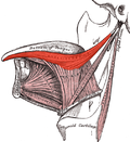
| m. hyoglossus | m. genioglossus | m. styloglossus |
The extraglossal muscles start on nearby structures, radiate to the tongue and move it as a whole. Extraglossal muscles include:
Palatoglossus muscle[edit | edit source]
- Beginning: edge of palatum molle
- Attachment: radix linguae
- Innervation: vagus nerve (X.)
- Function: pulls the radix linguae cranially
Genioglossus muscle[edit | edit source]
- Beginning: middle of the mandible ( spina musculi genioglossi ).
- Insertion: radiates fan-like into the tongue at the septum linguae , up to the aponeurosis linguae .
- Innervation: hypoglossal nerve (XII).
- Function: caudal bundles - pull the radix linguae forward, other bundles - pull the tongue to the floor of the mouth.
Styloglossus muscle[edit | edit source]
- Beginning: processus styloideus and ligamentum stylomandibulare .
- Attachment: on the sides of the tongue to the tip.
- Innervation: hypoglossal nerve (XII).
- Function: pulls the tip of the tongue back, the body and root back up.
Hyoglossus muscle[edit | edit source]
- Beginning: cornu majus of the tongue.
- Insertion: ventrocranial to the tongue (lateral to the previous muscle).
- Innervation: hypoglossal nerve (XII).
- Function: pulls the tongue back and down.
Intraglossal muscles[edit | edit source]
The intraglossal muscles begin and end in the tongue. They are arranged in 3 mutually perpendicular directions. Their movement changes the shape of the tongue.
The intraglossal muscles include:
Superior longitudinal muscle
- Origin and attachment : extends sagittally below the aponeurosis linguae (is connected to it).
- Innervation : hypoglossal nerve (XII).
- Function : shortens the language.
Inferior longitudinal muscle
- Beginning and attachment : it goes anteroposteriorly along the outer side of the genioglossus muscle (entwined with the bundles of the genioglossus muscle and the hyoglossus muscle).
- Innervation : hypoglossal nerve (XII).
- Function : shortens the language.
Musculus transversus linguae
- Beginning and attachment : it goes across from the septum linguae to the edges of the tongue between the bundles of the other muscles (extra- and intraglossal).
- Innervation : hypoglossal nerve (XII).
- Function : narrows the tongue.
Musculus verticalis linguae
- Beginning and attachment : dorsum linguae, attaches to the base of the tongue (goes between the bundles of other muscles).
- Innervation : hypoglossal nerve (XII).
- Function : flattens the tongue.
Vessels and nerves of the tongue[edit | edit source]
Arteries of the tongue[edit | edit source]
The artery of the tongue is a.lingualis (from the a.carotis externa) – it sends out rami dorsales linguae, arteria profunda linguae and arteria sublingualis.
Veins of the tongue[edit | edit source]
They lead to the vena lingualis and vena comitans nervi hypoglossi.
Lymphatic vessels[edit | edit source]
The lymphatic vessels of the tongue drain from the tip into the nodi lymphatici submentales, from the edge into the nodi lymphatici submandibulares, but most drain directly into the nodi lymphatici cervicales profundi.
Nerves of the tongue[edit | edit source]
Motor innervation – see muscles of the tongue.
Sensitive innervation is provided by the lingual nerve (from the 3rd branch of the trigeminal nerve), the facial nerve – apex, corpus; glossopharyngeal nerve (IX.) – radix; nervus laryngeus superior (from the vagus nerve (X.) – X.) – valleculae epiglotticae. The sensory innervation leads the gustatory component and initially its path is the same as the sensitive one, only from the first two thirds of the tongue then it leaves the lingual nerve via the chorda tympani and enters the facial nerve , where the taste fibers have their pseudounipolar cells in the ggl. geniculi.
Links[edit | edit source]
Related articles[edit | edit source]
External links[edit | edit source]
- Histologický preparát - jazyk
- HORKÝ, Drahomír – NOVÁKOVÁ, Květoslava. Morfologie orofaciálního systému pro studenty zubního lékařství [online] . 2. edition. 2011. Available from <http://mefanet.upol.cz/clanky.php?aid=58>. ISBN 978-80-244-2702-7.
References[edit | edit source]
- ČIHÁK, Radomír. Anatomie 2. 1. edition. Avicenum, 1988. pp. 488. ISBN 80-247-0143-X.
- JUNQUEIRA, L. Carlos – CARNEIRO, José – KELLEY, Robert O.. Basic histology. a LANGE medical book edition. Prentice Hall International (UK) Ltd., 1992. 488 pp. ISBN 0-8385-0587-2.
- KONRÁDOVÁ, Václava – UHLÍK, Jiří – VAJNER, Luděk. Funkční histologie. 2. edition. H & H, 2000. 291 pp. ISBN 80-86022-80-3.
- MARTÍNEK, Jindřich – VACEK, Zdeněk. Histologický atlas. 1. edition. Grada, 2009. ISBN 978-80-247-2393-8.


