Maxilla
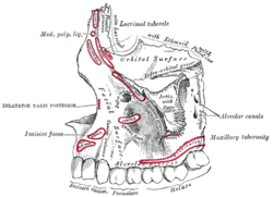
Maxilla (upper jaw) is the base of the facial part of the skull . It is a paired bone consisting of a body from which projections to the surrounding bones emerge.
Anatomical structures[edit | edit source]
Corpus maxillae[edit | edit source]
The body of the upper jaw is divided into a total of four surfaces:
- facies anterior – face surface
- contains the margo infraorbitalis as the lower border of the orbit
- foramen infraorbitale opens from the surface of the orbit
- caudally, it passes into the processus alveolaris, which is the base for the teeth, at the same time dependent on them
- he dental beds in which the teeth are stored are referred to as alveoli dentales
- facies infratemporalis – dorsal surface
- the rounding of the tuber maxillae is prominent here
- there are also numerous foramina alveolaria containing canals with the nerves and vascular supply of the posterior teeth
- facies orbitalis – forms part of the surface of the orbit and is in contact with the lacrimal, olfactory and cheekbones
- between the nearby ala major ossis sphenoidalis and facies orbitalis is the fissura orbitalis inferior, where it passes
- n. zygomaticus, n. infraorbitalis (II. trigeminal branch)
- a. infraorbitalis s v. ophthalmica inferior
- sulcus infraorbitalis, also begins here , which passes into a canal and opens as the foramen infraorbitale
- between the nearby ala major ossis sphenoidalis and facies orbitalis is the fissura orbitalis inferior, where it passes
- facies nasalis – rea forming part of the outer wall of the nose
- it contains formations created by the presence of the adjacent lacrimal bone as well as the hiatus maxillaris, leading to the sinus maxillaris, which forms one of the Paranasal sinuses
Processus frontalis[edit | edit source]
An anteriorly projecting projection in the cranial direction, where it connects with the frontal bone, nasal and lacrimal bones. At the same time, it looks into the Nasal cavity with its lateral surface and medially contains the edges for connecting concha nasalis media and inferior.
Processus zygomaticus[edit | edit source]
A similar, triangular and short process that joins the os zygomaticum in a suture.
Processus palatinus[edit | edit source]
The horizontal plate diverging medially from the maxilla merges with the symmetrical part of the second maxilla in the sutura palatina mediana. It also contains the spina nasalis anterior, which is located ventrally on the maxilla. The dorsal spina nasalis posterior is part of the os palatinum.
The front part of the maxilla, bearing dentes incisivi is called the premaxilla (os incisivum) and is important from the point of view of ontogenesis, when this part was separate and later fused with the maxilla. It leaves behind the canalis incisivus leading some nerves from the nasal cavity to the palate. thumb|200px|View of the Palate from below
Most common variants[edit | edit source]
Common are cleft defects, persistence of fusion of the premaxilla with the maxilla in the sutura incisiva, mouth of the canalis orbitalis in the maxillary sinus, or variations in the location of the mouth in the foramen infraorbitale.
Links[edit | edit source]
Related articles[edit | edit source]
Used literature[edit | edit source]
- ČIHÁK, Radomír. Anatomie I. 2. edition. Praha : Grada, 2001. 516 pp. pp. 163-166. ISBN 978-80-7169-970-5.


