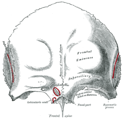
Frontal bone
Frontal bone is a pair-forming bone cranial vault. However, it fuses in adulthood and sometimes the fusion is visible as a metopic suture. It consists of three main sections.
Anatomical Structures[edit | edit source]
Squama frontalis[edit | edit source]
The scale of the frontal bone adjoins a pair of parietal bones dorsally in the coronal suture – the sutura coronalis.
- tubera frontalia - are paired bumps after the ossification centers, in which the scale is considerably curved;
- arcus superciliares - arcs located above the eyebrows;
- between the arches there is a glabella area in the middle;
- margo supraorbitalis borders the edge of the orbit, there are two incisions for vessels and nerves from the 1st branch of the n. trigeminus (n. ophthalmicus);
- incisura frontalis – medial notch, incisura supraorbitalis – lateral notch.
From the inside of the scale of the frontal bone, the imprints of the venous vessels (sulcus sinus sagittalis superioris) are visible, together with the impressions of the brain convolutions and vessels dura mater. In the center of the bone is a conspicuous crista frontalis where the dura mater is attached in vivo.
Partes orbitales[edit | edit source]
The parts embedded in the socket have visible imprints of the cerebral convolutions on the inner side of the brain. There are two dimples on the side of the orbit:
- fossa glandulae lacrimalis - laterally located pit for lacrimal glands,
- fossa trochlearis - medial fossa for the cartilaginous pulley over which the tendon of the eye muscle bends.
The partes orbitales continue medially as ceilings for the labyrinthi ethmoidales olfactory bones behind the passage of some vessels and nerves through small openings.
Pars nasalis[edit | edit source]
The nasal part of the frontal bone continues into the os nasale and proc. frontalis maxillae. Directly in the bone is a hollow opening lined with mucous nasal cavities, which is referred to as sinus frontalis' and belongs to the secondary nasal cavities. The cavity is divided asymmetrically by a septum and communicates through a paired passage with the nasal cavity (mostly together with cellulae ethmoidales anteriores of the olfactory bone). The sinus frontalis extends partially into the squama frontalis as well.
Most common variations[edit | edit source]
Metopism (sutura supranasalis) is a frequent variation that is caused by a persistent metopic suture from the fusion of the two bones. On normal bone, almost always only a trace of the suture remains. Other variations are a bony protrusion at the site of the cartilaginous pulley or an increased number of notches for formations on the margo supraorbitalis.
Links[edit | edit source]
Related Articles[edit | edit source]
References[edit | edit source]
- ČIHÁK, Radomír, et al. Anatomy I. 2. edition. Prague : Grada, 2001. 516 pp. pp. 155-158. ISBN 978-80-7169-970-5.


