Osteoid osteoma
Osteoid osteoma is a painful tumour up to 2 cm in diameter that occurs in people under 20 years of age in the cortical (superficial compaction) of long bones, especially the femur or tibia. It is usually located eccentrically in the diaphysis or metaphysis.
Osteoblastoma is mainly located in the pineal gland and does not sclerotize as much.
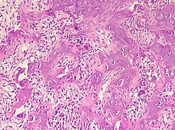
It consists of both an outer sclerotic rim (hyperostosis zone) and an inner so-called nidus (a tangle of fine osteoid lamellae, sometimes calcified, with osteoblasts perched on the surface of the lamellae, sometimes with osteoclasts present, the spaces between the lamellae are filled with a considerable cellular and vascular stroma). It may malignify into osteosarcoma.
Histology
Histological findings are similar to osteoblastoma.
Clinical picture
Manifested by nocturnal pain that subsides after antiplatelet agents (acylpyrine).
Diagnosis
X-ray examination shows a sclerotized bone deposit with spherical central lucency.
Therapy
It is treated by surgical removal of the nidus or radiofrequency ablation.
Links[edit | edit source]
Related Articles[edit | edit source]
Source[edit | edit source]
- PASTOR, Jan. Langenbeck's medical web page [online]. [cit. 18.04.2010]. <http://langenbeck.webs.com>.


