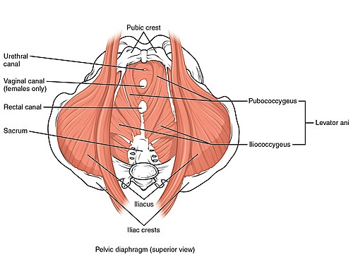
Pelvic floor muscles
The pelvic floor is made up of fibrous-muscular structures (diaphragm pelvis and diaphragma urogenitale). They attach to the bony base of the small pelvis, thereby closing the pelvic outlet and thus providing the necessary support for the pelvic organs.
Both membranes meet in the area of the centrum perineale, which is a key point for ensuring the integrity of the pelvic floor. Although these are structures with different functions and innervation, their mutual cooperation is necessary for the proper function of the sphincters (ensuring continence) and allowing the passage of the fetus through the birth canal (extreme dilation of the birth canal by relaxing the muscles of the pelvic floor).
Diaphragma pelvis[edit | edit source]
A flat funnel-shaped membrane that extends from the walls of the pelvis and attaches to the hiatus analis, where the anus passes.
It consists of the levator ani muscle and the coccygeus muscle. It extends from the walls of the small pelvis to the hiatus analis. Slot, between both mm. the levator ani form a passage called the hiatus urogenitalis, through which the urethra and (in women, together with the vagina) pass. Cranially, the muscles of the pelvic membrane are covered by the fascia diaphragmatis pelvis superior (a continuation of f. pelvis parietalis), caudally by the fascia diaphragmatis pelvis inferior.
Main function pelvic diaphragm is to support pelvic organs. All muscles of the pelvic diaphragm are innervated by direct branches from the sacral plexus (S3–S4).[1].
M. levator ani[edit | edit source]
A paired muscle that forms the ventrolateral part of the pelvis diaphragm. It consists of the pubococcygeus muscle (pars pubica), the iliococcygeus muscle (pars iliaca) and the puborectalis muscle.
- M. pubococcygeus
- It forms the ventral part of the pelvic floor. It begins in the common beginning at the pubic axis, (1 cm from the symphysis)[1] and in the course it divides into (m. pubovaginalis , m. puboperinealis and m. puboanalis).
- The muscle bundles cross each other during the course, forming a loop that forms an active support of the pelvic organs.
- The bundles are subsequently attached to various structures (reciprocal muscle, hiatus urogenitalis, lig. anococcygeum, up to the coccyx).
- M. iliococcygeus
- It forms the lateral part of the pelvic floor. It starts as a fibrous band on the arcus tendineus of the levatoris ani muscle and attaches to the lig. anococcygeum and coccyx margins.
- M. puborectalis
- It forms the lower border of the pelvic floor. It departs from the dorsal surface of the os pubis , runs laterally from the pubococcygeus muscle to the area of the anorectal junction, where it attaches to the bundles of the bilateral muscle. The attachment of the muscles creates a cuff surrounding the rectum, which pushes it ventrally and regulates stool continence by changing the contraction (relaxation of the muscle leads to defecation). At the same time, it affects the pelvic tilt and thus determines the value of the anorectal angle .
M. coccygeus[edit | edit source]
It forms the back part of the pelvic floor. It begins at the spina ischiadica and attaches to the area of the sacrococcygeal junction.
Urogenital Diaphragm[edit | edit source]
The fibrous part extends in the shape of a triangle, between the tubera ischiadica and the symphysis . It closes the hiatus urogenitalis through which the urethra passes (in women and the vagina) and in the area of the center of the perineum it connects with the diaphragma pelvis. Axial bodies and glands insist on the lower side.
In men, the muscular part consists of the transversus perinei profundus muscle and the variable transversus perinei superficialis muscle. In women, it is formed by the muscles of the sphincter urethrovaginalis and the compressor urethrae.
Superficial muscles of the perineum[edit | edit source]
They attach to the external genitalia and form the base of the perineum. They create two formations, according to which we divide them into the muscles of the urogenital and anal triangle.
The anal triangle consists of the sphincter ani externus muscle, the fibrous membrane and the lig. anococcygeum, which attaches the anal region of the rectum to the coccyx.
- The muscles of the urogenital triangle
-
- m. sphincter urethrae externus;
- m. ischiocavernosus (in men connected to the crus penis, participates in erection a ejaculation, in female connected to crus clitoridis);
- m. bulbospongiosus (in men the conclusion of micturition and ejaculation, in female contraction of the vestibule of the vagina, compression of the vestibular glands and erection of the clitoris).
Links[edit | edit source]
Related articles[edit | edit source]
References[edit | edit source]
- ČIHÁK, Radomír – GRIM, Miloš. Anatomie. 2. edition. Praha : Grada Publishing, 2002. 470 pp. vol. 1. ISBN 80-7169-970-5.
- HÁJEK, Zdeněk – ČECH, Evžen – MARŠÁL, Karel, et al. Obstetrics. 3. edition. Praha : Grada, 2014. 538 pp. ISBN 978-80-247-4529-9.

