Skin
The skin is the largest organ of the human body with a surface area of up to 2 m 2 , it makes up 5-9% of body weight.
The skin is composed of three main parts: epidermis (skin), corium (dermis) and tela subcutanea (subcutis - subcutaneous tissue).
Epidermis[edit | edit source]
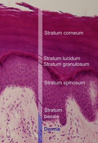
The epidermis is of ectodermal origin, it forms a stratified squamous keratinized epithelium. The average thickness is approximately 0.3-1.5 mm. The projections of the corium, the so-called papillae, extend into the lower border of the epidermis. Maturation of cells from the basal layer to the surface takes about 28 days, e.g. on the head this maturation takes 14 days. Cells from non-keratinized layers, keratinocytes , turn into scales, corneocytes , by keratinization .
We distinguish 5 layers according to cell type: stratum basale, stratum spinosum, stratum granulosum, stratum lucidum and stratum corneum.
Stratum basale (basal layer)
The stratum basale consists of one layer of palisade-like cubic-cylindrical keratinocyte cells and melanin granules , which form a cap above the nucleus. The cells are connected to each other by desmosomes, the connection to the basement membrane zone is via hemidesmosomes . Approximately 5% of cells called melanocytes form melanin. Melanocytes form numerous projections through which melanin is transported to the surrounding keratinocytes (1 melanocyte supplies approx. 30–40 keratinocytes). Melanocytes are not attached to neighboring keratinocytes by desmosomes, but are attached by hemidesmosomes to the basement membrane. Because of this, tumor cells (melanoma) metastasize quickly and easily.
Stratum spinosum
Together with the stratum basale, they form the stratum germinativum Malpighii. These are polygonal cells connected by desmosomes (they look like thorns under a microscope). Their intercellular spaces are filled with tissue fluid. The mechanical resistance of the skin is ensured by cytokeratin filaments.
Stratum granulosum
It contains one or more layers of flat cells. They have basophilic staining coarse grains of keratohyalin (keratin, trichohyalin, profillagrin,...), which are an intermediate product of keratinization. It protects the skin from the effect of substances from the environment by releasing granules of a phospholipid character by exocytosis into the intercellular spaces (Odland's bodies).
Stratum lucidum
The stratum lucidum is a thin layer of the epidermis containing 2-3 layers of cells. The nuclei are no longer stainable, the cytoplasm is homogeneous. In this layer, keratohyalin is transformed into granules of glycogen and eleidin. It forms an important barrier and is most prominently developed on the palms and soles.
Stratum corneum
It consists of several layers of anucleate, completely flattened cells, corneocytes. According to the thickness of this layer, we distinguish thick and thin type of epidermis. The strongest type is on the feet and palms. The stratum corneum is divided into two parts - the stratum conjunctum (the lower compact layer) and the stratum disjunctum (the upper peeling layer).
Langerhans cells[edit | edit source]
These are dendritic cells, penetrating all layers of the epidermis. They arise in the bone marrow , have the ability to present antigens to lymphocytes ("antigen presenting cells"). Their number is different, increases during inflammation, decreases due to UV radiation . They have long projections and light chromophobe cytoplasm. They can be distinguished from keratinocytes by the presence of ATPase; protein S 100+.
Merkel cells[edit | edit source]
They are mechanoreceptors, located in the stratum basale and the outer epithelial sheath of the hair follicle. They have oval nuclei, light cytoplasm, their peptide granules can be demonstrated immunohistochemically.
Corium[edit | edit source]
Corium is of mesenchymal origin. It is made up of fibrous fibers, ground substance, nerves, blood vessels and cells.
It has 2 distinct layers stratum papillare and stratum reticulare.
Stratum papillare
This part extends towards the epidermis in the form of papillae. It forms a sparse collagen fiber with many cells and elastic fibers. There are free sensitive nerve endings, nerve bodies − Meissner's, Ruffini's...
Stratum reticulare
The stratum reticulare can be found under the papillary layer in the form of a dense mesh of collagen and elastic fibers. There are fewer cells, but it contains lobules of fat cells and Vater-Pacini corpuscles.
Construction of coria[edit | edit source]
Fibrous component
It consists of 4 types of fibers:
- collagen - skin strength, oriented (skin cleavage lines)
- elastic – supportive, surrounding the adnexa , strength and flexibility
- reticulin - fine
- anchoring fibrils – connecting the basement membrane to the collagen fibers of the dermis
Extracellular matrix
Cellular elements
Cell elements include fibroblasts, histiocytes, mast cells, and lymphocytes.
Blood vessels
We divide into two systems - superficial (subpapillary) and deep. Among them are the rami communicantes.
Blood vessels disappear
They start in the papillae and also form 2 systems.
Nerves
Within sensitive nerves, these are simple fibrils (free nerve endings) or specialized endings. These endings include the Ruffini corpuscle, Meissner corpuscle or Vater-Pacini corpuscle. The Ruffini corpuscle is surrounded by perineurium and collagen fibers. Meissner's corpuscles are oval in shape, larger than Ruffini's, and are found on the tips of the fingers, palms, soles, lips, and tongue. It is an accumulation of Schwann cells and collagen fibers that bind the basement membrane to the epidermis. The Vater-Pacini corpuscle is 1-3 mm in size and has an oval shape. It registers pressure sensations, occurs not only in the skin, but also in the walls of internal organs, blood vessels and tendons. Vegetative nerves have the functions of glands, they are capable of vasoconstriction and dilation. They also cause spasms of the arrectores pilorum muscle.
Subcutaneous bodies[edit | edit source]
The hypodermis is of mesenchymal origin. There are ligaments, blood vessels, nerves, nerve endings and sweat glands. Thinnest on the eyelids, thickest on the buttocks, abdomen and thighs.
Skin adnexa[edit | edit source]
More detailed information can be found on the Skin adnexa page .
Links[edit | edit source]
related articles
- Physiology of the skin
- Skin development
- Skin function
- Phototypes| Pigment | Pigmentation disorders
- Thick skin - HE
- Histologie: Thick-type skin (histological specimen) | Axilla/histological specimen | Exercise: Skin system
Source
- BENEŠ, Jiří. Study materials [online]. ©2007. [cit. 4.11.2010]. <http://jirben2.chytrak.cz/>.
Literature
- MARTÍNEK, Jindřich – VACEK, Zdeněk. Histological atlas. 1. edition. Prague : Grada, 2009. 136 pp. ISBN 978-80-247-2393-8.
- ŠTORK, Jiří. Dermatovenerology. 1. edition. Prague : Galén, Karolinum, 2008. ISBN 978-80-7262-371-6.
- JIRSOVÁ, Zuzana. Skin [lecture for subject Histology and Embryology, specialization General Medicine, 1. LF UK]. Prague. 2011.
External links


