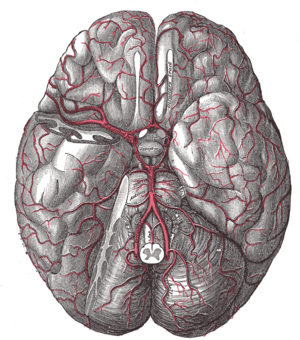
Subarachnoid haemorrhage
In subarachnoid hemorrhage, blood leaks into the liquor pathways between the arachnoid and pia mater. It is a massive bleeding from the arterial basin.
The causes of subarachnoid haemorrhage[edit | edit source]
It most often occurs due to a rupture of an aneurysm (60%), in the area of the circle of Willis, especially on the a. communicans anterior or posterior, often with increased blood pressure (physical exertion, coitus, agitation, defecation, etc.). Other causes may be trauma, vascular malformations such as arteriovenous and cavernous malformations, anticoagulant therapy, bleeding disorders, hypertension, amyloid angiopathy, primary vasculopathy. In addition, idiopathic (cryptogenic) SAKs occur. Traumatic SAKs are then often associated with contusion.
Clinical picture[edit | edit source]
The headache appears within seconds and can be very intense. It is localized bilaterally, sometimes with a maximum occipitally. Initially, there may be a brief disturbance of consciousness. The pain is further accompanied by nausea, vomiting, photophobia and phonophobia. A meningeal syndrome develops within minutes to hours. Patients are often disoriented, confused, some patients are somnolent to soporific, sometimes, on the contrary, psychomotor restlessness, aggressiveness, negativism may dominate. When subarachnoid haemorrage propagates intracerebrally, focal symptomatology develops. The patient's condition is evaluated with the Hunt and Hess scale - see vascular diseases of the brain.
CAVE!!! In some cases, symptoms may be less intensely expressed and mimic more of a cervicocranial syndrome, so in unclear cases we always indicate brain CT scan and LP (lumbar puncture).
Diagnostics[edit | edit source]
The diagnosis is made by a CT scan. Approximately 5% of CT scans do not show subarachnoid haemorrhage in the first 24 hours, so if subarachnoid haemorrhage is still suspected, we indicate a lymph node examination. The typical liquor finding is oxyhemoglobin on spectrophotometric examination.
CAVE!!! Liquor must be processed within 1 hour of collection. We also find elevated protein and thousands to hundreds of thousands of erythrocytes on cytology, phagocytosis of erythrocytes from day 3-4, and maximum bilirubin on spectrophotometry.
Treatment[edit | edit source]
If subarachnoid haemorrhage is proven, we refer the patient to neurosurgery for cerebral panangiography, which should be performed within 72 hours of the onset of the symptoms due to the risk of vasospasm. If an aneurysm is found and the H+H score is within 3, surgery is indicated - either clipping the aneurysm neck or filling the aneurysm cavity with a detachable coil - coiling. Bed rest is essential (hospitalisation always), symptomatic treatment (analgesics, antiemetics, correction of hypertension). Vasospasm can be suppressed with calcium ion blockers.
If the aneurysm is not proven to be the cause, the patient is treated conservatively - opioids for pain, mucolytics and laxatives, and after 3-6 weeks a control panangiography is indicated.
Bleeding from an aneurysm[edit | edit source]
An aneurysm is a circumscribed enlargement of a cerebral artery. The aneurysm is caused by long-term effect of blood pressure on the weakened vascular wall (congenital defects, atherosclerosis, mycotic infections, trauma, etc.). The bulge thins the vascular wall, which ruptures with a sudden increase in blood pressure (exertion, defecation, coitus, agitation, bending over). Aneurysms are most often found in the region of the Circle of Willis, mainly on the arteria carotis interna, a. communicans ant., a. cerebri media).
Bleeding from cerebral aneurysms is relatively common in the Czech Republic (about 600 SAC cases per year), unfortunately with a very high lethality (40% of patients die after the first bleed). The aneurysms themselves (especially smaller ones) are clinically mute until rupture and bleeding occur.
Clinical picture[edit | edit source]
Manifestations are sudden, dramatic and rapidly progressive. The initial symptom is an intense headache (sharp, not yet recognized), nausea, vomiting, increase in systemic pressure due to Cushing's reflex, photophobia, impaired consciousness, and there may be localized neurological findings depending on the location of the aneurysm, epileptic seizures, meningism, increased body temperature, lesions of the cranial nerves (III and VI), etc.
Diagnosis[edit | edit source]
To establish the diagnosis of subarachnoid haemorrhage, a CT scan is necessary (evidence of blood in the liquor spaces, intracerebral hematoma), which can also roughly outline the source of bleeding. This is then clarified by cerebral panangiography, which we try to perform as soon as possible after the diagnosis of subarachnoid haemorrhage, and always in at least two projections. In patients without intracranial hypertension in whom CT was not sufficiently conclusive, we perform CSF collection and examination (to detect the presence of erythrocytes or xanthochromine).
The therapeutic approach is based on the Hunt and Hess classification:
- grade 0 - non-bleeding aneurysm, without symptoms,
- grade I - headache, neck opposition, mild meningeal syndrome,
- grade II - headache, neck opposition, lesions of the cranial nerves, more pronounced meningeal syndrome,
- grade III - dullness or confusion, mild lesion
- stage IV - stupor, decerebral rigidity, vegetative disorders,
- stage V - deep coma, decerebral rigidity.
Therapy[edit | edit source]
- Conservative therapy
- for deferred operations
- if coagula block the liquor pathways, we introduce external ventricular drainage
- Surgical therapy
- closure of the aneurysm neck while maintaining blood flow through other vessels
- the procedure is a prevention of rerupture, it does not repair the damage caused by the previous subarachnoid haemorrhage
- early operations are among the most complex neurosurgical operations
- Endovascular methods
- coiling - obliteration of the aneurysm with metal spirals
- spirals have shape memory and coil themselves around the aneurysm, a coagulum forms on them and the entrance to the aneurysm is re-epithelialized
- it is done angiographically, under an X-ray lamp, little burden on the patient
- limitations of coiling - reachability of the aneurysm by catheterization, ratio of the neck to the aneurysm (when it is large, the coils leak)
- if the aneurysm has a wide neck, a stent is usually implanted first and coils are inserted through it to prevent leakage
- coiling - obliteration of the aneurysm with metal spirals
Arteriovenous malformations[edit | edit source]
An A-V malformation (AVM) is a congenital convoluted artery and vein that communicate directly with each other and between which no capillary system has been formed. As a result of the defect, the resistance of the capillaries is negligible, resulting in increased blood flow in the malformed vessels. As a result, other areas are less well supplied with blood and are subject to ischaemia (steal phenomenon).
Clinically, it is manifested by haemorrhage (70%) not only in subarachnoid haemorrhage but also intracerebral. Ischaemia of less blooded parts leads to localized manifestations (e.g. epileptic seizures).
For the basic diagnosis we use CT and MRI, the source of bleeding is then identified by PAG.
Therapy[edit | edit source]
Malformations tend to be extensive and often difficult to access surgically. Unless they are very acute cases, we choose the "watch and scan" method, which assesses the condition of individual vessels and their progression over time. We then perform the procedure on arteries that could be at risk.The operation consists of closing the feeding arteries by bipolar coagulation (the procedure takes many hours and is difficult). The alternative is embolization of the arteries endovascularly or the use of a Leksell gamma knife.
Cavernous hemangioma[edit | edit source]
Cavernous haemagngioma is a special kind of AVM. A cavernoma is a circumscribed, small vascular mass in brain tissue that does not have wide feeding arteries. For this reason, it is not readily visible on PAG, so we choose MRI for diagnosis.
Bleeding is not extensive, but it is quite frequent.
Differential diagnosis[edit | edit source]
- Subdural haemorrhage
- Epidural bleeding
- Migraine
- Acute cervicocranial syndrome
- Meningitis
Links[edit | edit source]
Related articles[edit | edit source]
- Treatment of intracranial aneurysm
- Craniocerebral Trauma
- Blood vessels of the brain
- Stroke
- Circuit of Willis
- Aneurysms
- Arteriovenous malformation
- Cavernous malformation
Used literature[edit | edit source]
- NEVŠÍMALOVÁ, Soňa, Evžen RŮŽIČKA a Jiří TICHÝ. Neurologie. 1. vydání. Praha : Galén, 2005. s. 163-170. ISBN 80-7262-160-2
- AMBLER, Zdeněk. Základy neurologie. 6. vydání. Praha : Galén, 2006. s. 171-181. ISBN 80-7262-433-4
- ZEMAN, Miroslav, et al. Speciální chirurgie. 2. vydání. Praha : Galén, 2004. 575 s. ISBN 80-7262-260-9
Sources[edit | edit source]
- BENEŠ, Jiří. Studijní materiály [online]. ©2007. [cit. 2009]. <http://www.jirben.wz.cz/>


