The large intestine (specimen)
Large intestine B4[edit | edit source]
Overview[edit | edit source]
Specimen 1[edit | edit source]
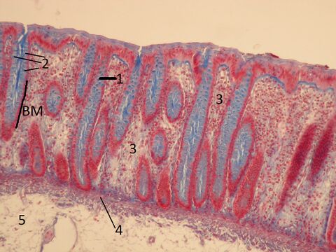
Name: Large intestine (colon) AZAN
Description: The epithelium of the large intestine is single-layered cylindrical. When stained with AZAN, the nuclei are colored red and the mucin in goblet cells is colored blue. The epithelial cells form simple tubular glands (Lieberkühn's crypts) in the lamina propria (loose collagenous tissue).
1 - epithelium lining the crypt 2 - individual goblet cells 3 - lamina propria mucosae, loose collagenous tissue 4 - lamina muscularis mucosae 5 - tunica submucosa
Specimen 2[edit | edit source]
Name: Large intestine (colon) (HE)
Description: The mucosa of the large intestine - in a cross section, sections of Lieberkuhn's crypts can be seen. The lighter cells are goblet cells. Mucin does not stain with regular processing. The spaces between the crypts are filled with loose collagenous tissue with a large number of free cells (lymphocytes, plasma cells, etc.).
1 - crypt in cross-section 2 - lamina propria 3 - accumulation of mucinous granules in goblet cells 4 - colonocytes
Specimen 3[edit | edit source]
Title: Large intestine (colon) PAS
Description: PAS reaction stains polysaccharides, glycoproteins, and proteoglycans pink (magenta). In the preparation, PAS reaction stains mucin in goblet cells in the epithelium and slightly less in the interstitial tissue in the connective tissue.
1 - crypt 2 - lamina propria mucosae 3 - lamina muscularis mucosae 4 - submucosal layer BM - basal membrane
Specimen 4[edit | edit source]
Title: Large intestine (HE)
Description: Large intestine - slight enlargement. In addition to the mucosal layer (lamina epithelialis - single-layered cylindrical epithelium; lamina propria mucosae - thin collagenous connective tissue; lamina muscularis mucosae - smooth muscle), the submucosal layer is also visible, which is also made up of thin connective tissue, but is much denser compared to the connective tissue in the lamina propria, that is, it contains more fibers and fewer cells.
Specimen 5[edit | edit source]
Title: Colon, submucosal layer - submucosal plexus
Description: The submucosal plexus (small ganglion in the center of the field of view) is located in the thin collagenous connective tissue of the submucosal layer just below the lamina muscularis mucosae. It contains multipolar neurons surrounded by satellite cells and connected by nerve fibers.
Specimen 6[edit | edit source]
Title: Large intestine; PAS and Hematoxylin staining
Description: PAS stains polysaccharides, proteoglycans and glycoproteins in the tissue (cyclamen pink - magenta). In the preparation, PAS is positive for mucin in goblet cells (single-cell intraepithelial glands).


