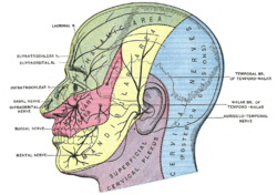
Trigeminal nerve
The trigeminal nerve is the fifth cranial nerve. It is the strongest of all cranial nerves. It has both a sensitive and a motor component. There are also parasympathetic fibers running somewhere, but they only attach to it. These fibers come from VII (n. facialis) and IX (n. glossopharyngeus).
The trigeminal nerve is also an afferent part of important reflexes (eg masseter, corneal).
Inervation[edit | edit source]
It sensitively innervates the entire face, the oral cavity: hard and soft palate up to the isthmus faucium, the front two thirds of the tongue, all the teeth and the nasal cavity, the entire contents of the orbit, most of the dura mater, part of the auricle.
It motorically innervates the chewing muscles (m. masseter, m. temporalis, m. pterygoideus medialis, m. pterygoideus lateralis), m. mylohyoideus, anterior belly m. digastricus.
Cores[edit | edit source]
- nucleus motorius nervi trigemini - the only motor nucleus (also as ncl. masticatorius), fibers run only in V3, medial from the sensitive nucleus of the trigeminal nerve
- nucleus pontinus (principalis) - the somatosensitive fibers from the ggl. trigeminale end here,
- nucleus mesencephalicus – proprioception from muscles, gums, teeth and oculomotor muscles,
- nucleus spinalis - "somatosensitive" fibers from ggl. trigeminale + from ganglia n. VII., IX., X..
Tribe Progress[edit | edit source]
The exit of the nerve is from the ventral edge of the pontus, then the nerve continues to the depression of the tip of the pyramid – impressio trigemini. A ganglion trigeminale (semilunare, Gasseri) is placed between the two sheets of dura mater. Here, pseudounipolar cells send dendrites to the periphery and axons to the brain to nuclei. It has three main branches: n. ophthalmicus, n. maxillaris and n. mandibularis.
Ophthalmic nerve[edit | edit source]
The most medial branch of the ggl. trigeminal
About 2.5 cm long and continues forward in the lateral wall of the cavernous sinus
Innervation[edit | edit source]
- Somatosensitively
- in the eye environment - orbit and the adjacent periosteum, eyeball, lacrimal gland, skin of the forehead and upper eyelids, conjunctiva,
- in the nose area - the skin of the back and tip of the nose, the mucous membrane of the olfactory labyrinths, the sphenoid sinus and the ventral part of the nasal cavity.
Tribe Progress[edit | edit source]
From the ggl. trigeminale, it goes ventrally in the lateral wall of the sinus cavernosus, where it runs under the oculomotor and trochlearis nerves and enters the fissura orbitalis superior. Here it divides and progresses towards the orbital (the part above the annulus tendineus communis - n. lacrimalis et n. frontalis, the part inside the annulus - n. nasociliaris).
Branch[edit | edit source]
- Connections with sympathetic knitting on the surface a. carotis interna.
- Connections to n. III., IV., VI.
- Ramus tentorii (meninges).
- Nervus nasociliaris - enters through the annulus tendineus, lateral to the n. opticus, continues through it up to the inner wall of the orbit
- nervus ethmoidalis posterior - sensitively innervates the walls of the sinus sphenoidalis and cellulae ethmoidales
- nervus ethmoidalis anterior - up into the cranial cavity, down and through the lamina cribrosa ossis ethmoidalis penetrates into the nasal cavity, where it innervates the mucous membrane in the upper and front part
- ramus nasalis externus
- nervus infratrochlearis - under the trochlea m. obliquus superior bulbi to the inner corner of the eye and splits into branches of the rami palpebrales
- nervi ciliares longi
- ramus communicans cum ganglio ciliary (radix nasociliaris ganglii ciliaris).
- Nervus frontalis - enters through the fissura orbitalis superior above the anulus tendineus communis, under the ceiling of the orbit above the m. levator palpebrae superioris, it further divides into
- nervus supratrochlearis - more medial, innervates the conjunctiva, the skin of the upper eyelid, the glabella, the skin of the root of the nose and the inner corner of the eye
- nervus supraorbitalis- more lateral and coarser, between the levator palpebrae superioris muscle and the ceiling of the orbit and passes through the incisura supraorbitalis to the forehead area, is divided into: ramus medialis and ramus lateralis, innervates the upper eyelash , the conjunctiva of the eye, the skin of the forehead and head up to the region of the parotid, the branches and the sinus frontalis
- Nervus lacrimalis - continues to the side wall of the orbit, where it is along the upper edge of the rectus lateralis bulbi muscle together with the a. lacrimalis under the outer edge of the orbital roof to the outer corner, where it receives from V2n. zygomaticus and with it "parasympathetic" fibers for the lacrimal gland, sensitive innervation of the conjunctiva of the eye and the skin of the lateral part of the upper eyelash
- r. communicans lacrimalis nervi zygomatici - parasympathetic fibers from ggl. pterygopalatinum and through the zygomatic nerve reach the lacrimal gland
Maxillary nerve[edit | edit source]
The middle of the three branches of V2
It exits the skull through the foramen rotundum, enters the pterygopalatine fossa and continues to the inferior orbital fissure.
Innervation[edit | edit source]
- Somatosensitively
- hard diaper at canalis rotundus,
- in the area of the maxilla - maxilla, upper teeth, sinus maxillaris, mucous membrane of the posterior two thirds of the cavitas nasi, palate, isthmus faucium, skin in the area from the eye slits to the mouth, mucous membrane of the upper half of the cheeks with nasopharynx and Eustachian tubes.
- Parasympathetic
- fibers for the innervation of the glandula lacrimalis, enter through the zygomatic n. into the "n. lacrimalis" (see above),
- glands of the nasal mucosa, salivary glands of the palate, upper lip and upper half of the face.
Tribe Progress[edit | edit source]
From the semilunar ganglion, it passes down the lateral wall of the sinus cavernosus to the canalis rotundus to the fossa pterygopalatina. Here it branches out.
Branch[edit | edit source]
- Ramus meningeus - coverings of the brain
- Nervi pterygopalatini (rr. ganglionares) – nerves passing through the pterygopalatine ganglion without connection, innervate sensitive areas in the mucous membrane of the nasal cavity, palate and pharynx, parasympathetic fibers for the lacrimal gland
- Nervus infraorbitalis - from the pterygopalatine fossa goes through the fissura orbitalis inferior into the orbit, on the lower part of which it passes into the canalis infraorbitalisand emerges on the ventral surface of the maxilla in the foramen infraorbitale
- rami alveolares superiores posteriores - plexus dentalis superior
- ramus alveolaris superior medius,
- rami alveolares superiores anteriores
- rami cutanei
- rami palpebrales inferiores
- rami nasales externi
- rami labiales superiores
- rami nasales posteriores superiores laterales - to the foramen sphenopalatinum and from there to the nasal cavity
- rami nasales posteriores superiores mediales
- rami nasales posteriores inferiores
- n. palatinus major - to the mucous membrane of the hard palate
- n. pharyngeus
Ganglion pterygopalatinum - the largest parasympathetic ganglion in the pterygopalatine fossa, receives parasympathetic fibers via the major petrosus nerve, sympathetic fibers from the plexus caroticus internus via the petrosus profundus nerve
- Nervus zygomaticus - from the pterygopalatine fossa through the fissura orbitalis inferior into the orbit, along the external wall to the foramen zygomaticorbitale in the os zygomaticum, here it divides into two branches
- nervus zygomaticofacialis - skin in the cheek region above the os zygomaticum
- nervus zygomaticotemporalis - to the skin of the temple, infraorbitalis
- r. communicans lacrimalis nervi zygomatici – parasympathetic connection from the orbit to n. lacrimalis for the lacrimal gland.
Nervus mandibularis[edit | edit source]
The lateral branch, containing only the motor fibers from the ventral trunk
Area of innervation[edit | edit source]
- Somatosensitively from the dorsal trunk
- land lower jaw including skin, mucous membrane, teeth and gums,
- landscape of the sleeping area.
- Branchiomotoric – 4 masticatory muscles + 2 supragyoid
- m. mylohyoideus, venter anterior musculi digastrici,
- fibers from nuclei n. VII for m. tensor tympani; n. IX for m. tensor veli palatini.
Tribe Progress[edit | edit source]
Exit from ggl. trigeminale, passing through the foramen ovale under the skull base to the fossa infratemporalis and branches here.
Ganglion oticum - is a parasympathetic ganglion, which receives its preganglionic parasympathetic through the branches from n. IX. After the connection, the fibers lead to the glandula parotis and also innervate vessels and glands in the area of the sensitive innervation of the n. mandibularis. Fibers for the tensor tympani and tensor veli palatini come "motorically". The sympathetic fibers come together with the n. petrosus minor and with the sympathetic weaving around the a. meningea media. After passing through the ganglion, fibers are added to innervate vessels, smooth muscle, and glands.
Branch[edit | edit source]
- Ramus meningeus - sensitive innervation dura mater.
- Rami musculares - motor, ventral trunk
- masseteric nerve
- nervi temporales profundi
- nervus pterygoideus lateralis et medialis
- nervus tensoris veli palatini and nervus tensoris tympani,
- mylohyoid nerve.
- Nervus buccalis - a sensitive branch in the fossa infratemporalis to the face, cross ductus parotideus
- Nervus auriculotemporalis - a sensitive nerve running under the skull base dorsally, forming a loop for a. meningea media and continuing in front of the auricle up to the skin of the auricle and sleepscapes
- rami communicantes cum ganglio otico
- rami membranae tympani
- rami communicantes cum nervo faciali
- rami parotidei
- rami articulares,
- nervus meatus acustici externi,
- nervi auriculares anteriores
- rami temporales superficiales
- Nervus lingualis - the first strong branch going between the pterygoideus lateralis et medialis m. in the caudal direction to the hypoglossus m. to the mucous membrane of the tongue (front 2/3), floor of the mouth, palatine tonsil, isthmus faucium and gingiva
- connection with chorda tympani, connection with n. facialis, which brings preganglionic parasympathetic fibers to ganglion submandibulare a sublinguale, located at n. lingualis, from n .lingualis to the facial nerve, sensitive fibers from the tongue go and to the lingual nerve, parasympathetic fibers from the facial nerve go
- rami communicantes ad nervo hypoglosso
- rami isthmi faucium (rr. tonsilares),
- nervus sublingualis - innervates the mucous membrane of the oral cavity under the tongue and the gums of the front teeth of the lower molars
- rami linguales.
- Nervus alveolaris inferior - the second strong branch with a sensitive and motor component.
After giving off the motor fibers, it continues to the canalis mandibulae to innervate the adjacent structures
- nervus mylohyoideus – sensitive and motor innervation of the mylohyoideus muscle, venter anterior digastric muscle
- plexus dentalis inferior – sensitive, parasympathetic and sympathetic fibres,
- nervus mentalis – final section - rr. mentales, labiales, gingivales
Clinic[edit | edit source]
- Infection herpes zoster often spreads in the innervation area of the ophthalmic nerve.
- Unilateral palsy of the trigeminal nerve results in sensory loss in the area innervated by the terminal branches of the nerve, loss of motor fibers is relatively rare.
- Trigeminal neuralgia is a designation for severe irritating pain in areas innervated by sensitive fibers of the supraorbital nerve, the infraorbital nerve and the mental nerve. The most common cause is compression of the superior cerebellar nerve at the point of transition between the CNS and the PNS, where the sheaths of oligodenocytes and Schwann cells touch.
Investigation[edit | edit source]
Examination of the trigeminal nerve is most often performed using two reflexes:
- Masseter reflex - induced by hitting the lower teeth with a spatula inserted in the mouth, thereby stretching the masseter muscle tendon and causing it to contract.
- Corneal reflex - caused by a light touch of the cornea with the aim of contracting the orbicularis oculi muscle = blinking.
Links[edit | edit source]
Related Articles[edit | edit source]
References[edit | edit source]
- PETROVICKÝ, Pavel – DRUGA, Rastislav. Systematická, topografická a klinická anatomie : VIII. periferní nervový systém. 1. edition. Nakladatelství Karolinum, 1997. ISBN 80-7184-108-0.
- ČIHÁK, Radomír – GRIM, Miloš. Anatomy 3. 2nd, upr. a dopl edition. Grada, 2004. 673 pp. vol. 3. pp. 477-487. ISBN 80-247-1132-X.
- KACHLÍK, David. Hlavové nervy 1. díl : Přednáška [online]. ©2012. [cit. 2012-01-26]. <http://old.lf3.cuni.cz/anatomie/Hlavovenervy1.zip>.


