Vagina
Vagina[edit | edit source]
Description[edit | edit source]
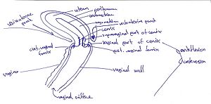
Vagina is a musculomembranous tube, 7-9cm long, extending from the cervix of uterus to the vestibule of vagina. It is usually collapsed: its anterior and posterior walls are in contact except from the superior part where the cervix keeps them apart. The vagina is compressed by 4 muscles: Pubovaginalis, External urethral sphincter, Urethrovaginalis sphincter, Bulbospongiosus.
It has 4 fornices, that form around the vaginal part of the cervix:
- Posterior vaginal fornix. The deepest part and closely related to rectouterine pouch
- Anterior vaginal fornix
- Lateral (left) vaginal fornix
- Lateral (right) vaginal fornix
It serves for:
- As an excretory canal for cervical fluid
- Forms the inferior part of the pelvic canal
- Receives penis and ejaculate during sexual intercourse
- It connects the cervical canal (from the isthmus to the external os of uterus) to the vestibule of vagina.
Vasculature & Innervation[edit | edit source]
- Arterial supply: by the uterine arteries for superior part and middle and inferior parts are supplied by the vaginal and internal pudendal arteries.
- Venous drainage: by the vaginal venous plexus which communicates with the uterine, vesical and rectal venous plexuses.
- Lymphatic drainage: superiorly -> int.+ext. iliac nodes, middle -> int. iliac nodes, inferiorly -> sacral + common iliac nodes
- Innervation: derived from the uterovaginal plexus. Inferior part of vagina is innervated by the pudendal nerve.
Topographic relations[edit | edit source]
- Anteriorly: fundus of bladder + urethra
- Laterally: levator ani, visceral pelvic fascia + ureters
- Posteriorly (inferior to superior): anal canal, rectum, rectouterine pouch
External female genital organs[edit | edit source]
The external female organs include the following:
- labia majora
- labia minora (they have anterior and posterior commissures and form a frenulum on the posterior one)
- the clitoris
- the hymen
- the vestibule of the vagina
- the bulb of vestibule
- the greater vestibular gland
- the external urethral orifice (with openings of ducts of paraurethral glands).
The hymen is a thin anular fold of mucous membrane surrounding the lumen of the vaginal orifice, and after its rupture, its remnants are found as hymen caruncles. The skin folds that form external borders of vagina (and together with mons pubis and they form a triangle).
The thin skin folds surround vestibule of vagina, that divide anteriorly into 2 folds that unite over the clitoris to form the prepuce of clitoris.
The clitoris originates on 2 limbs, the crura. The crura are covered by the ischiocavernosus muscles and originate from the lower rami of the pubic bones. The body of clitoris is formed by the union of the 2 crura just below the pubic symphysis, and it terminates anteriorly as glans of clitoris. The greater vestibular glands (mucus-secreting) are located on either side of the vestibule of vagina. Directly anterior to them, the bulbs of vestibule (erectile tissues) are located, which are covered by 1 bulbospongiosus muscle each.
Links[edit | edit source]
Related articles[edit | edit source]
Bibliography[edit | edit source]
- MOORE, Keith L – DALLEY, Arthur F. Clinically Oriented Anatomy. 5. edition. Lippincott Williams & Wilkins, 2005. ISBN 0781736390.


