Uterus
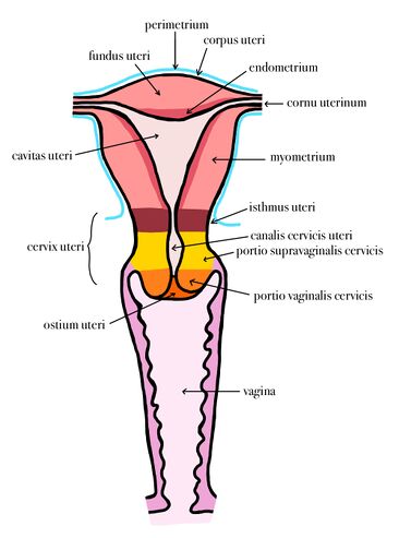
Uterus (womb) is a hollow muscular organ with a thick wall located in the small pelvis of a woman. It is pear-shaped and slightly anteroposteriorly sheathed. The dimensions of the uterus vary according to the stage of woman's life (in a woman who has not yet given birth = nullipara, the dimensions are 8 x 5 x 2-3 cm (length x width x depth), in a woman who has already given birth = multipara, the dimensions are slightly larger).
Parts of the uterus:
- fundus uteri - arcuate part above the body of the uterus
- corpus uteri - uterine body;
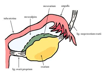
- cornu dextra + sinistra - horns of the uterus, openings of the fallopian tubes
- cervix uteri - cervix; divided into portio vaginalis (cervix) and portio supravaginalis
- isthmus uteri - a short segment connecting the corpus and cervix.
Borders (= margo) and surfaces (= facies) can also be seen on the uterus:
- facies vesicalis (= anterior) - anterior surface facing the bladder
- facies intestinalis (= posterior) - posterior surface facing the villi of the small intestine
- margo dextra + sinistra - right and left border of the uterus
Anatomy of the uterus[edit | edit source]
The uterus is located in a small pelvis between the bladder and the rectum. It is anteroposteriorly sheathed (in adulthood) and occupies a typical position:
- anteflexio uteri – the longitudinal axis of the uterus forms an obtuse angle (160-170°) with the axis of the cervix, this angle is open to the front;
- anteversio uteri – the angle between the uterus and the vagina (70-100°).
Structure of the uterine wall (histological structure of the uterus)[edit | edit source]
The uterine wall is about 15 mm thick and is made up of three basic layers:
- the endometrium;
- the myometrium;
- the perimetrium (serosa or adventitia).
Endometrium[edit | edit source]
The surface of the endometrium is covered by a simple columnar epithelium. Beneath the epithelial layer is the lamina propria, which is composed of simple tubular glands (glandulae uterinae). The stroma is highly cellular - rich in fibroblasts. Functionally, the endometrium is divided into 2 basic layers:
- stratum basalis – this layer remains the same, it does not change during the menstrual cycle. It contains a base of glands, more collagenous and reticular fibers;
- stratum functionalis – during the menstrual cycle, hormones cause changes in the structure of this layer and its separation. The bodies of the glands penetrate this portion. The hormone estrogen from the base of the glands (stratum basalis) is involved in regeneration.
The endometrium is supplied with blood from the arteriae arcuatae, which are divided into 2 sets of arteries, depending on which part of the endometrium they supply:
- straight arteries – supplying stratum basalis;
- spiral arteries (arteriae spirales) – supplying stratum functionalis.
Myometrium[edit | edit source]
The myometrium is a thick layer of smooth muscle divided into 4 (some sources say 3) loosely demarcated layers:
- stratum submucosum with a predominance of longitudinal bundles;
- stratum vasculare with predominance of longitudinal bundles and rich vasculature;
- stratum supravasculare with a predominance of circularly oriented bundles;
- stratum subserosum again with a predominance of longitudinally oriented bundles.
In addition to muscle fibres, the myometrium contains an admixture of connective tissue, vascular and lymphatic supply and autonomic nerves.
During pregnancy, this layer undergoes great changes - there is both hypertrophy, but also hyperplasia of muscle cells.
Perimetrium and parametrium[edit | edit source]
The superior and posterior surface of the uterus is covered by a serous membrane - the perimetrium, which partly extends to the anterior wall of the uterine body. On the sides it transitions into the lig. latum uteri. It is formed by the mesothelium. The subserosal connective tissue transitions into the fixation apparatus of the uterus - parametrium. The lower section of the uterus (anterior wall of the cervix) is covered by the adventitia.
Blood supply and lymphatic drainage of the uterus[edit | edit source]
Arterial supply:
- a. uterina - from a. iliaca interna
Venous drainage:
- plexus venosus uterovaginalis - drains into vv. uterinae and subsequently ends up in v. iliaca interna
Lymphatic drainage
- nodi lymphatici lumbales - drains lymph from the fundus and corpus uteri
- nodi lymphatici iliaci interni - drains lymph from the corpus, isthmus and cervix uteri
- nodi lymphatici sacrales - drains lymph from the isthmus and cervix uteri
- nodi lymphatici inguinales superficiales - drains lymph from the margines and cornua uteri
Changes in the uterus during the menstrual cycle[edit | edit source]
During the menstrual cycle, depending on the level of sex hormones, changes take place in the uterine endometrium. These changes mainly involve the zona functionalis, which proliferates during the menstrual cycle, simple tubular glands branch and grow, and secretion is produced due to progesterone stimulation (from the corpus luteum in the ovary). This prepares the uterine lining to receive the fertilized egg. However, if fertilization has not occurred, the corpus luteum disappears, progesterone production ceases and a new menstrual cycle begins - menstruation and with it the loss of the zona functionalis. The zona basalis is thinner, contains the basal parts of the uterine glands and is not subject to changes during the menstrual cycle.
Changes in the uterus during a woman's life[edit | edit source]
During life, the shape of the uterus, its size and proportions change. All of this depends on the level of estrogens.
Changes in the uterus depending on the period of life:
- typus uteri infantilis = resting period - in children, the uterine body is smaller than the cervix (2:1 ratio);
- typus uteri pubertalis – during puberty, the level of estrogens rises sharply and the uterine body responds to this by increasing (1:1 ratio);
- typus uteri adultae –the uterine body gradually grows until the ratio between it and the cervix increases to 2:1. Gradually, its position is adjusted to anteflexion and anteversion;
- typus uteri senilis – as a result of insufficient estrogen stimulation, the uterine body shrinks again and the uterus resembles the typus uteri pubertalis.
Changes in the uterus during pregnancy:
- the uterus grows depending on the growth of the fetus;
- takes on a spherical shape;
Links[edit | edit source]
Related articles[edit | edit source]
Literature[edit | edit source]
- ČIHÁK, Radomír. Anatomie II. 2. edition. Praha : Grada, 2001. 488 pp. ISBN ISBN 80-247-0143-X.
- JUNQUEIRA, L. Carlos – CARNEIRO, José – KELLEY, Robert O.. Základy histologie. 7. edition. H & H, 1997. 502 pp. a LANGE medical book; ISBN 80-85787-37-7.
- ROB, Lukáš – MARTAN, Alois – CITTERBART, Karel. Gynekologie. 2. edition. Praha : Galén, 2008. 390 pp. ISBN 978-80-7262-501-7.
- ČECH, Evžen – HÁJEK, Zdeněk – MARŠÁL, Karel, et al. Porodnictví. 2. edition. Praha : Grada, 2006. 544 pp. ISBN 80-247-1303-9.
- LÜLLMANN-RAUCH, Renate. Histologie. - edition. Grada Publishing a.s., 2012. 556 pp. ISBN 9788024737294.


