Congenital defects of the nervous system: Difference between revisions


Vlckovaanna2 (talk | contribs) |
Vlckovaanna2 (talk | contribs) |
||
| Line 165: | Line 165: | ||
==[[Dandyova-Walkerova malformace]]== | ==[[Dandyova-Walkerova malformace]]== | ||
__notoc__ | __notoc__ | ||
| type = article | | type = article | ||
| surname1 = Osenbach | | surname1 = Osenbach | ||
| Line 260: | Line 261: | ||
Therapy consists in removing the obstruction, and hydrocephalus is treated by introducing a shunt. | Therapy consists in removing the obstruction, and hydrocephalus is treated by introducing a shunt. | ||
<noinclude> | <noinclude> | ||
== Links == | |||
=== Related articles === | |||
* [[CNS malformations]] | |||
* [[Congenital defects of the nervous system]] | |||
=== External links === | |||
* http://www.priznaky-projevy.cz/geneticke-nemoci/449-dandy-walker-syndrom-dandy-walkerova-malformace-priznaky-projevy-symptomy | |||
=== Literature used=== | |||
* {{Cite | |||
| type = book | |||
| isbn = 80-7345-072-0 | |||
| surname1 = SAMEŠ | |||
| name1 = M | |||
| group = yes | |||
| title = Neurochirurgie | |||
| edition = 1 | |||
| location = Praha | |||
| publisher = Jessenius Maxdorf | |||
| year = 2005 | |||
}} | |||
* {{Cite | |||
| type = article | |||
| surname1 = OSENBACH | |||
| name1 = Richard K. | |||
| surname2 = MENEZES | |||
| name2 = Arnold H. | |||
| article = Diagnosis and Management of the Dandy-Walker Malformation: 30 Years of Experience | |||
| journal = Pediatric Neurosurgery | |||
| url = https://www.karger.com/Article/Abstract/120660 | |||
| year = 1992 | |||
| volume = 18 | |||
| number = 4 | |||
| pages = 179-189 | |||
| issn = - | |||
| doi = 10.1159/000120660 | |||
}} | |||
=== References === | |||
{{cite | |||
| type = article | |||
| surname1 = Osenbach | |||
| name1 = R K | |||
| surname2 = Menezes | |||
| name2 = A H | |||
| article = Diagnosis and management of the Dandy-Walker malformation: 30 years of experience | |||
| journal = Pediatr Neurosurg | |||
| year = 1992 | |||
| number = 4 | |||
| volume = 18 | |||
| pages = 179-89 | |||
| url = https://www.ncbi.nlm.nih.gov/pubmed/1472430 | |||
| issn = 1016-2291 | |||
}} | |||
{{Cite | |||
| type = book | |||
| isbn = 978-1-4557-5980-4 | |||
| surname1 = Marcdante | |||
| name1 = Karen J | |||
| title = Nelson essentials of pediatrics | |||
| edition = 7 | |||
| year = 2015 | |||
| range = 0 | |||
}} | |||
{{Cite | |||
| type = article | |||
| surname1 = Can | |||
| name1 = Serdar Suleyman | |||
| surname2 = Karakaş Uğurlu | |||
| name2 = Görkem | |||
| surname3 = Cakmak | |||
| name3 = Selcen | |||
| article = Dandy walker variant and bipolar I disorder with graphomania | |||
| journal = Psychiatry Investig | |||
| year = 2014 | |||
| number = 3 | |||
| volume = 11 | |||
| pages = 336-9 | |||
| url = https://www.ncbi.nlm.nih.gov/pmc/articles/PMC4124195/?tool=pubmed | |||
| issn = 1738-3684 | |||
| doi = 10.4306/pi.2014.11.3.336 | |||
}} | |||
{{Cite | |||
| type = article | |||
| surname1 = Pandurangi | |||
| name1 = Swapna | |||
| surname2 = Pandurangi | |||
| name2 = Aditya | |||
| surname3 = Matkar | |||
| name3 = Abhay | |||
| group = yes | |||
| article = Psychiatric manifestations associated with mega cisterna magna | |||
| journal = J Neuropsychiatry Clin Neurosci | |||
| year = 2014 | |||
| number = 2 | |||
| volume = 26 | |||
| pages = 169-71 | |||
| url = https://www.ncbi.nlm.nih.gov/pubmed/24763763 | |||
| issn = 0895-0172 (print), 1545-7222 | |||
}} | |||
</noinclude> | |||
[[Category:Neurology]] | |||
[[Category:Neurosurgery]] | |||
[[Category:Pediatrics]] | |||
== Links == | == Links == | ||
=== Related articles === | === Related articles === | ||
Revision as of 15:22, 16 December 2022
Arachnoid cyst
An arachnoid cyst is a well-defined cystic, congenital collection of cerebrospinal fluid (it arises on the basis of doubling of the arachnoid layer). It is often an accidental finding on a CT scan or MRI scan. It is important for a differentia diagnosis of (tumors).[1] [2]
Symptoms and auxiliary tests
In the CT image (resp MRI, possibly with the intrathecal application of contrast) arachnoid cyst has a density, resp. cerebrospinal fluid intensity (hypodense). Most common locations:
- in the middle pit
- on convexity
- in the posterior cranial pit (cisterna magna)
- in the cerebellopontine angle
- at the chiasm level
- in the chamber
Expansive behaviour depends on its communication with the surrounding cerebrospinal fluid spaces. The problem occurs if the communication has a valve character (intracranial hypertension syndrome) occurs.
Arachnoid cysts can be asymptomatic, otherwise, they often cause symptoms in early childhood (increased intracranial pressure, epileptic seizures, gradually developing focal neurological symptoms, sudden worsening of the condition during cyst bleeding.[1][2]
- CT of arachnoid cysts of the temporal lobe

Sagittal plane

Frontal plane
Transverse plane


Treatment
An accidental finding and an asymptomatic cyst are not treated or followed up. For expansive behaviour and clinical manifestations, surgery is chosen (shunt surrounding cerebrospinal fluid, ie microsurgical / endoscopic fenestration of the cyst into the normal cerebrospinal fluid). A shunt formed between the arachnoid cyst (most often in the case of the sulcus lateralis cyst) and the peritoneal cavity, the so-called cystoperitoneal shunt, is also possible. Marsupialization (removal of the cyst with the capsule) is rarely performed.[1][2]

Chiari malformation
Chiari malformation (Arnold-Chiari malformation) is a congenital CNS anomaly. It is a dystopia of the cerebellum and medulla oblongata into the spinal canal, which is clinically manifested by hydrocephalus. We distinguish four types of rhombencephalon abnormalities (cerebellum, pons, oblongata):
- Type 1 - herniation of the cerebellar tonsil under the foramen magnum, IV. the chamber is stored normally
- Type 2 - usually the co-presence of myelomeningocele
- Type 3 - severe dislocation of structures in the posterior pit, often associated with suboccipital encephalomeningocele; usually incompatible with life
- Type 4 - cerebellar hypoplasia without herniation.

Clinical picture
Clinically, the defect is manifested mainly by headache, weakened grip and spasticity of DK.
Diagnosis
Native X-ray, MRI.
Therapy
The main problem is hydrocephalus – decompression of the craniospinal junction, short-circuit drainage operations.
Craniostenosis

Craniostenosis is caused by premature adhesion of sutures. The earlier it occurs, the more severe its effects. The shape of the head depends on the order in which the sutures close - as a suture grows, the skull stops growing perpendicular to the suture.
Normal development of the skull and sutures
- at birth, the skull has one lamina, and the diploe is formed at 4 years of age
- large fontanelle is 4x2.5 cm at birth, closes at 1.5-2.5 years
- the small one closes at 2-3 months
- the growth of the sutures is prevented by the proliferation of fibroblasts along the suture line
- CI - cephalic index - percentage ratio of head width to length - 60-70% in children, 70-90% in adults
- lengthening of the head decreases CI
Dividing
- primary - due to a developmental disorder of unclear etiology;
- secondary - in microcephaly and after drainage hydrocephalus.
Diagnosis
- clinically - palpation of fontanelles, X-ray, CT
- at high pressure, gyral impressions are seen on the skull bones - the so-called skull of wrought silver
- scintigraphy - open suture receives Tc, closed not
Types
- scaphocephaly - sagittal suture fusion, the head is narrow, resembles a ship's keel
- brachycephaly - affects the coronal suture - flat forehead, greater distance between orbits, etc.
Therapy
- many surgical methods, indications are intracranial hypertension or cosmetic
- early surgery can ensure normal psychomotor development
Meningocele

Meningocele (spina bifida cystica) is a split of the vertebral arch, from which emerges a sac made up of soft spinal cords (the dura mater usually ends at the neck of the sac). The content is cerebrospinal fluid, 1/3 of children are neurologically affected.
'Menigomyelocele (spina bifida aperta) is a variant of meningocele that occurs a little more often and is less favorable. In addition to the packages, there is also spinal cord in the hernial sac.
Therapy
On admission, place the child on the stomach and exclude pressure on the meningomyelocele. In case of perforation, apply ATB and cover the bearing with a mule. We operate within 24 hours. In complete motor paraplegia and sphincter lesions, this cannot be improved by any surgery, but surgery is in order to prevent infection.
Neurofibromatosis
Neurofibromatosis is a relatively common AD hereditary disease (1:2,500–4,000 newborns) based on cells derived from the neural bar. It is manifested by abnormal growth of CNS and PNS support cells (Schwann's bb. etc.) with a pronounced predisposition to the formation of benign and malignant tumors[3].
The disease belongs to hereditary tumor syndromes, it occurs in two forms. Familial forms of these syndromes arise as a result of a congenital mutation of tumor-suppressor genes. A certain percentage of these syndromes arise as a result of new mutations[4]. Molecular-genetic analysis is available for definitive confirmation of this diagnosis.
Forms
There are two basic forms of this disease, which differ both in the cause (mutation of different genes) and in the consequences (different clinical picture).
Neurofibromatosis – Type 1
Neurofibromatosis – type 1 (NF-1, also called morbus von Recklinghausen or peripheral type of neurofibromatosis) is conditioned by a mutation of the NF1 gene on chromosome 17 (17q11.2)[5]. It is a tumor-suppressor gene whose product (neurofibromin) is part of the intracellular signaling cascade associated with THE RAS-kinase.
![]() It is necessary to distinguish morbus Recklinghausen, which is synonymous with primary hyperparathyroidism.
It is necessary to distinguish morbus Recklinghausen, which is synonymous with primary hyperparathyroidism.
Clinical manifestations of this form include:
- So-called "café-au-lait spots" (spots of the color of "white coffee", in 90% appear up to 5 years of age).[4]
- Neurofibromas (multiple tumorous nodules; cutaneous, subcutaneous and plexiform; mainly in the axillae and groin).
- Lisch nodules (hamartomy iris).[4][5]
- Increased risk of developing various cancers: CNS gliomas (optics gliomas), neurofibrosarcoma, rhabdomyosarcoma, pheochromocytoma, leukemia, etc.[4]
- Involvement of the musculoskeletal system (subperiosteal neurofibromas – causing hypertrophy of the bone, its thinning and pathological fractures, scoliosis, congenital dysplasia of the tibia)[3].
- Intellectual impairment, epilepsy or stenosis of the renal artery.[4][5]
Neurofibromatosis – Type 2
Neurofibromatosis – type 2 (NF-2, also called MISME syndrome or central type neurofibromatosis) is conditioned by mutation of the NF2 gene on chromosome 22 (22q12.2)[5]. It is also a tumor-suppressor gene whose product (neurofibromin 2, also called merlin or schwannomine) affects intercellular contacts. Central neurofibromatosis is generally rarer than the peripheral type, but overall it is associated with higher morbidity and mortality of affected individuals.[4] About half of NF-2 cases are caused by a new mutation.[5]
Clinical manifestations of this form include:
- CNS tumors: meningeomas, astrocytomas, ependymomas, spinal root schwanoms, retinal hamartoms (MISME syndrome = Multiple Inherited Schwannomas, Meningiomas, and Ependymomas).
- Bilateral vestibular schwannoma is particularly typical.
- Also in this form we find "café-au-lait" spots, but not Lisch's nodules.[4]
Therapy
- Causal therapy does not exist.
- Dispensary of patients with a proven diagnosis of neurofibromatosis is suitable.
- Surgical interventions indicated in case of nerve oppression / obstruction in the GIT form, possibly from a cosmetic point of view.[3]
- Neurosurgical interventions in CNS involvement; possible use of stereotactic neurosurgery (Leksell's gamma knife).
Dandyova-Walkerova malformace
 | type = article
 | surname1 = Osenbach
 | name1 = R K
 | surname2 = Menezes
 | name2 = A H
 | article = Diagnosis and management of the Dandy-Walker malformation: 30 years of experience
 | journal = Pediatr Neurosurg
 | year = 1992
 | number = 4
 | volume = 18
 | pages = 179-89
 | url = https://www.ncbi.nlm.nih.gov/pubmed/1472430
 | issn = 1016-2291
}} </ref> | prognóza = | MKN = Template:MKN | MeSH ID = | OMIM = 220200 | orphanet = | MedlinePlus = | Medscape = Template:Medscape }}
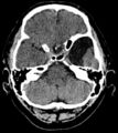
Dandy-Walker syndrome is a rare disease, affecting the development of the brain. It manifests with a triad of symptoms
- complete or partial agenesis of vermis cerebellum
- cystic dilatation of the fourth ventricle of brain
- enlargement of the posterior cranial fossa[6]
Enlargement of the posterior cranial fossa may be associated with membranous atrasia of the apertures of the fourth ventricle, leading to pathological accumulation of cerebrospinal fluid in the ventricles. These abnormalities These abnormalities most often result in problems with movement, coordination but also with intellect. Psychiatric illness may also occur[7]. Affected individulas may survive up to their second decade of life.
Malformations
In most cases, individuals with Dandy-Walker syndrome develop symptoms of abnormal brain development during the first year of life. Clinically, it is manifested in most children by the accumulation of cerebrospinal fluid in the brain - hydrocephalus, which can cause macrocephaly. Mental disability is often present[8] , although some individuals also have a normal intellect. Children often have delayed development, especially in the field of motor skills (crawling, walking, coordination of movements). Muscle stiffness and paralysis - spastic paraplegia - may occur. Older children experience symptoms of increased intracranial pressure such as irritability, vomiting, convulsions and nystagmys. Less frequently other cerebral malformations are present such as agenesis of the corpus callosum connecting the right and left hemispheres, occipital encephalocele or abnormal clefts in the brain - schizencephaly and gyrification disorders. These brain defects are associated with more or less severe signs and symptoms. Dandy-Walker syndrome can also include heart defects, malformations of the urogenital tract, limbs and face. A milder form of the disease is the Dandy – Walker variant, which includes hypoplasia of the cerebellar vermis without agenesis, mild or no enlargement of the posterior fossa and fourth ventricle, without hydrocephalus.
Genetics
Mutations in certain genes occur in individuals affected by Dandy-Walker syndrome, but these mutations account for only a small number of all known cases. Dandy-Walker syndrome is also associated with chromosomal abnormalities on most chromosomes.'' It is most common in people with trisomy 18, but can also occur with trisomy 13, 21 or 9. Dandy-Walker syndrome has also been reported in fetuses with triploidy, a fatal condition in which individuals have an extra complete set of chromosomes in each cell. Dandy-Walker syndrome can also be caused by mutations of specific genes (FOXC1 contains instructions for the preparation of a protein that regulates the activity of other genes, ZIC1 and ZIC4 act as transcriptional activators and participate in CNS organogenesis). Brain malformations associated with Dandy-Walker syndrome can occur also in isolation, and may not be associated with a genetic disease, with the cause being often unknown.
Environmental influence
Dandy-Walker syndrome can be caused by environmental factors that affect the fetus before birth. For example exposure of the fetus to rubella, toxoplasmosis or due to substances that causes damage to the fetus, (teratogens). A more frequent occurrence of fetuses with Dandy-Walker syndrome also occurs in the fetuses of mothers who suffer from diabetes.
Diagnosis and incidences
Prenatal diagnosis is possible by ultrasound examination and performing amniocentesis to examine the karyotype of the fetus. It is estimated that Dandy-Walker syndrome occurs in 1:25000 to 1:30000 newborns. The occurrence of cases of Dandy-Walker syndrome is rare and does not have a clear pattern of inheritance. Only ``immediate relatives of people with Dandy-Walker syndrome have an ``increased risk of developing the disease compared to the general population.
Therapy
Therapy consists in removing the obstruction, and hydrocephalus is treated by introducing a shunt.
Links
Related articles
External links
Literature used
- SAMEŠ, M. Neurochirurgie. 1. edition. Praha : Jessenius Maxdorf, 2005. ISBN 80-7345-072-0.
- OSENBACH, Richard K. – MENEZES, Arnold H.. Diagnosis and Management of the Dandy-Walker Malformation: 30 Years of Experience. Pediatric Neurosurgery [online]. 1992, vol. 18, no. 4, p. 179-189, Available from <https://www.karger.com/Article/Abstract/120660>. DOI: 10.1159/000120660.
References
OSENBACH, R K – MENEZES, A H. Diagnosis and management of the Dandy-Walker malformation: 30 years of experience. Pediatr Neurosurg [online]. 1992, vol. 18, no. 4, p. 179-89, Available from <https://www.ncbi.nlm.nih.gov/pubmed/1472430>. ISSN 1016-2291.
MARCDANTE, Karen J. Nelson essentials of pediatrics. 7. edition. 2015. 0 pp. ISBN 978-1-4557-5980-4.
CAN, Serdar Suleyman – KARAKAŞ UĞURLU, Görkem – CAKMAK, Selcen. Dandy walker variant and bipolar I disorder with graphomania. Psychiatry Investig [online]. 2014, vol. 11, no. 3, p. 336-9, Available from <https://www.ncbi.nlm.nih.gov/pmc/articles/PMC4124195/?tool=pubmed>. ISSN 1738-3684. DOI: 10.4306/pi.2014.11.3.336.
PANDURANGI, Swapna – PANDURANGI, Aditya – MATKAR, Abhay. Psychiatric manifestations associated with mega cisterna magna. J Neuropsychiatry Clin Neurosci [online]. 2014, vol. 26, no. 2, p. 169-71, Available from <https://www.ncbi.nlm.nih.gov/pubmed/24763763>. ISSN 0895-0172 (print), 1545-7222.
Odkazy
Související články
Externí odkazy
Použitá literatura
Reference
- ↑ Jump up to: a b c SEIDL, Zdeněk – OBENBERGER, Jiří. Neurologie pro studium i praxi. 1. edition. Praha : Grada Publishing, 2004. ISBN 80-247-0623-7.
- ↑ Jump up to: a b c SAMEŠ, M, et al. Neurochirurgie. 1. edition. Praha : Jessenius Maxdorf, 2005. ISBN 80-7345-072-0.
- ↑ Jump up to: a b c DUNGL, P., et al. Ortopedie. 1. vydání. Praha : Grada Publishing, 2005. ISBN 80-247-0550-8.
- ↑ Jump up to: a b c d e f g KLEIBL, Zdeněk a Jan NOVOTNÝ. Hereditární nádorové syndromy. 1. vydání. Praha : Triton, 2003. 31 s. ISBN 80-7254-357-1.
- ↑ Jump up to: a b c d e FIRTH, Helen V., Jane A. HURST a Judith G. HALL. Oxford desk reference: clinical genetics. 1. vydání. Oxford : Oxford University Press, 2005. 708 s. ISBN 9780192628961.
- ↑ MARCDANTE, Karen J. Nelson essentials of pediatrics. 7. edition. 2015. 0 pp. ISBN 978-1-4557-5980-4.
- ↑ CAN, Serdar Suleyman – KARAKAŞ UĞURLU, Görkem – CAKMAK, Selcen. Dandy walker variant and bipolar I disorder with graphomania. Psychiatry Investig [online]. 2014, vol. 11, no. 3, p. 336-9, Available from <https://www.ncbi.nlm.nih.gov/pmc/articles/PMC4124195/?tool=pubmed>. ISSN 1738-3684. DOI: 10.4306/pi.2014.11.3.336.
- ↑ PANDURANGI, Swapna – PANDURANGI, Aditya – MATKAR, Abhay. Psychiatric manifestations associated with mega cisterna magna. J Neuropsychiatry Clin Neurosci [online]. 2014, vol. 26, no. 2, p. 169-71, Available from <https://www.ncbi.nlm.nih.gov/pubmed/24763763>. ISSN 0895-0172 (print), 1545-7222.
Kategorie:Neurologie
Kategorie:Neurochirurgie
Kategorie:Pediatrie
Dětská mozková obrna
Dětská mozková obrna (DMO, angl. cerebral palsy) je trvalá neprogredující porucha hybnosti provázená abnormálním svalovým napětím a abnormální posturou. Jedná se o nenakažlivé, nedědičné onemocnění, které vzniká na podkladě jednorázového poškození mozkové tkáně (nejčastěji hypoxií). Je charakteristické poruchou vývoje motorických oblastí mozku nebo jejich jiným poškozením v raném stádiu vývoje. DMO je následkem prenatální, postnatální nebo raně postnatálního poškození vyvíjejícího se mozku.
K poruše hybnosti se mnohdy připojuje epilepsie, poruchy citlivosti, smyslů (např. zrakové postižení) a vnímání, poruchy učení, kognice, komunikace, chování nebo mentální retardace. DMO je neurovývojovou poruchou, jejíž projevy se v průběhu vývoje většinou mění.
DMO patří do skupiny vývojových onemocnění, protože vzniká na základě širokého spektra abnormalit vyvíjejícího se CNS. Různé etiologie působící na různá vývojová stadia mohou vést ke stejnému klinickému obrazu a naopak obdobná etiologie může vyvolat odlišné následky.
Léčba DMO je komplexní, multioborová a dlouhodobá. Jejím cílem není vyléčení nebo dosažení normálního stavu, ale zvětšení funkčnosti, zlepšení schopností a udržování zdraví ve smyslu lokomoce, kognitivního vývoje, sociální integrace a nezávislosti. Úspěch terapie závisí na její včasnosti a intenzitě.
![]()
Klasifikace
- podle anatomické topografie postižení:
- mono-, hemi-, di-, kvadruparetická forma;
- podle patofyziologického typu hybné poruchy:
- spastické formy a nespastické formy: s dyskinezami, dystoniemi, hypotonií, ataxií.[1]
Epidemiologie
Incidence DMO je asi 2:1000 živě narozených dětí. Riziko roste nepřímo úměrně s gestačním stářím při porodu.[2]
Etiologie

Hybnou poruchu při dětské mozkové obrně způsobuje postižení supraspinálních hybných center, kortikospinálních traktů, segmentálních spinálních okruhů a muskuloskeletárního systému.[1]
Může vzniknout během jednoho ze čtyř následujících období:
- V těhotenství (prenatální etiologie) – intrauterinní infekce, léky nebo placentární dysfunkce;
- Při porodu (perinatální etiologie) – hlavně u vícečetných těhotenství, hypoxie, hemoragie, hypoglykémie, meningitida;
- V prvních měsících života dítěte (postnatální etiologie) – trauma, encefalopatie, encefalitida;
- Popř. velmi nízká porodní hmotnost.
V prvním a druhém trimestru vznikají poruchy vývoje CNS. Na začátku třetího trimestru to je periventrikulární leukomalácie (PVL) a intraventrikulární hemoragie (IVH). Ke konci třetího trimestru vznikají léze kortikální, subkortikální a léze hluboké mozkové šedi.[1]
- Bilaterální spastické a dyskinetické formy – hypoxicko-ischemická léze mozku.
- Diparéza – periventrikulární leukomalacie u předčasně narozených dětí.
- Spastická hemiparéza zralých novorozenců – infarkt a. cerebri media, periventrikulární glióza.
- Spastická hemiparéza nezralých novorozenců – periventrikulární porencefalie v důsledku intraventrikulárního krvácení.
- Dyskinetická forma – hypoxicko-ischemické poškození v oblasti thalamu a bazálních ganglií (děti po 32. týdnu těhotenství).
- Atetoidní forma – neonatální hyperbilirubinémie s jádrovým ikterem (velmi vzácná).
- Ataktická forma – většinou neznámá příčina, ev. strukturální porucha mozečku.[2]
Klinické příznaky

- forma: spastická, dyskinetická nebo ataktická;
- distribuce: bilaterální – diparéza, kvarduparéza, unilaterální – hemiparéza;
- míra postižení: škála GMFCS I – V (Gross Motor Function Classification System) – funkční test pro hodnocení míry a následných změn v hrubé motorice pomocí standardních volních pohybů (volní hybnost, chůze a sed); MACS (The Manual Ability Classification System) – test manuálních schopností;
- komorbidity: epilepsie, mentální retardace, smyslové poruchy.[1]
- Spasticita: abnormálně zvýšený svalový tonus, zvýšené svalové reflexy, pozitivní pyramidové jevy, abnormální pohyby a držení (pes equinus, vnitřní rotace a addukce v kyčlích, pronace a flexe předloktí), vznik kontraktur.
- Dystonie: abnormální trvalé svalové kontraktury, které vedou k abnormálnímu dystonnímu držení a abnormálním pohybům (flexe, pronace zápěstí s nataženými prsty nebo torzí trupu).
- Atetóza: generalizované, nekoordinované, přehnané, mimovolní hyperkinetické pohyby, normální nebo snížený svalový tonus.
- Ataxie: dysmetrie nebo intenční tremor horních končetin, ataxie chůze a stoje – postižení dolních končetin a trupu (stoj a chůze o široké bazi, kolíbavá chůze).[2]
Spastické formy DMO
Diparetická forma (někdy označována jako paraparetická)
- Nejčastější (1/3 postižených).
- Symetrické postižení obou dolních končetin (slabší, méně vyvinuté).
- Nápadný nepoměr mezi vzrůstem trupu a dolních končetin.
- Svalová hypertonie, zkraty svalů – vadné držení dolních končetin i pánve.
- Může být lehká až střední mentální retardace.
Hemiparetická forma
- Druhá nejčastější.
- Postižené končetiny slabší a zpravidla kratší ve srovnání s druhostrannými.
- Skoro vždy více postižena horní končetina.
- Typické postižení charakterizováno paží přitaženou k trupu, je pokrčena až úplně ohnutá v lokti, předloktí otočeno hřbetní stranou vzhůru, ruka ohnuta směrem do dlaně a uchýlena směrem k malíkové straně.
- Častý je výskyt epilepsie, případně může být i senzitivní deficit a mentální retardace.
Kvadruparetická forma
- Těžší forma diparetické formy.
- Obrnou postiženy všechny 4 končetiny.
- Přítomna je i epilepsie a mentální retardace či poruchy učení.
Dyskineticko-ataktické formy DMO
- Cca 10–15 % případů DMO.
- Epilepsie je u těchto forem vzácná.
- Převládá-li choreo-atetóza, bývá normální inteligence, ale slovní vyjadřování ztěžuje těžká dysartrie.
- Převládá-li dystonie nebo ataxie, častěji je přítomna i mentální retardace.
Onemocnění je často kombinací více forem DMO.
Diagnostika
- DMO je klinická diagnóza; definitivně stanovena je až po 3. roce věku (při nezralosti CNS jsou projevy nespecifické).
- Vyšetření k objasnění etiologie:
- UZ mozku, MR mozku;
- koagulace (protein C a S, homocystein, APC rezistence) – při prokázaném infarktu;
- EEG; vyšetření zraku a sluchu;
- genetické vyšetření – u lissencefalie.[2]
Diferenciální diagnostika
Pro DMO svědčí hlavně stabilní klinický nález bez progrese postižení. Pro stanovení rozsahu je důležité zobrazovací vyšetření (CT, MRI). Progrese postižení ukazuje spíše na neurodegenerativní či neurometabolické postižení. Pomalu rostoucí tumor CNS.
Terapie
DMO je choroba, která se nedá vyléčit. Dá se však pomocí léčby dosáhnout zlepšení podmínek a životních možností dítěte, což většinou příznivě ovlivňuje kvalitu jeho života. Lékařské pokroky posledních let a hlavně pokroky v léčbě postižených s DMO vedly k tomu, že dnes již mnozí z těch, kteří byli včas a správně léčeni, mohou vést téměř normální život. Ani dnes ještě neexistuje nějaká standardní léčba, která by byla u všech pacientů dostatečně účinná. Lékař, který léčí pacienta s DMO, je víceméně závislý na řadě specializovaných odborníků, s jejichž pomocí nejprve správně určí a stanoví individuální poruchy a pak jim přizpůsobí celý terapeutický program. V současné době probíhá také několik klinických studií na aplikaci kmenových buněk z pupečníkové krve.
Léčebný plán může zahrnovat:
- Rehabilitaci – zlepšení kontroly lokomoce i posturálního držení pomocí stimulačních a facilitačních technik (např. Vojtovou metodou), nejprve odborníky, posléze instruovanými rodiči.
- Antiepileptika.
- Myorelaxans u spastických forem.
- Dlahy pro svalovou nerovnováhu.
- Operační léčbu – ortopedické korekce (mezi 6. a 10. rokem), například u diparetické formy selektivní dorzální rizotomie (částečné přetětí zadních míšních kořenů ve výši L2).
- Protetické pomůcky.
- Aplikaci botulotoxinu.
- Speciální péči a výchovu od předškolního věku.
Odkazy
Související články
Externí odkazy
Použitá literatura
- ↑ Jump up to: a b c d
- ↑ Jump up to: a b c d
Kategorie:Neurologie Kategorie:Pediatrie
Odkazy
Související články
Zdroj
Reference
Kategorie:Neurologie
Kategorie:Embryologie
Kategorie:Genetika
