Stomach: Difference between revisions
No edit summary |
Khyativishu (talk | contribs) |
||
| (3 intermediate revisions by 3 users not shown) | |||
| Line 1: | Line 1: | ||
==Basic anatomy of the stomach== | ==Basic anatomy of the stomach== | ||
'''The stomach''' (''ventriculus, gaster, stomachos'') is a muscular sac located in the abdominal cavity under the left [[Diaphragm|diaphragmatic]] vault. It extends upward under the rib cage into the ''regio hypochondriaca sinistra'' (the region of the abdomen below the [[diaphragm]] at the cartilage of the lower [[ribs]]), from there the stomach goes down to the right into the ''regio epigastrica'' (between the right and left rib arches). | [[File:Stomach..jpg|thumb|410x410px|Stomach]] | ||
'''The stomach''' (''ventriculus, gaster, stomachos'') is a muscular sac located in the abdominal cavity under the left [[Diaphragm|diaphragmatic]] vault. It extends upward under the rib cage into the ''regio hypochondriaca sinistra'' (the region of the abdomen below the [[diaphragm]] at the cartilage of the lower [[ribs]]), from there the stomach goes down to the right into the ''regio epigastrica'' (between the right and left rib arches). | |||
*'''''curvatura major''''' – curvature of the left edge, curving downwards to the left; | It has '''the shape''' of a curved pouch with a left convex and a right concave edge (curvature): | ||
*'''''curvatura minor''''' – curvature of the right edge, concave upwards to the right; | |||
*'''''curvatura major''''' (''greater curvature'')– curvature of the left edge, curving downwards to the left; | |||
*'''''curvatura minor''''' (''lesser curvature'')– curvature of the right edge, concave upwards to the right; | |||
*'''''cardia''''' (comb) - the mouth of the [[oesophagus]] from above into the stomach at the curvature minor; | *'''''cardia''''' (comb) - the mouth of the [[oesophagus]] from above into the stomach at the curvature minor; | ||
*'''''pylorus''''' (gatekeeper) - a narrowed place, adjacent to the stomach [[duodenum]]. | *'''''pylorus''''' (gatekeeper) - a narrowed place, adjacent to the stomach [[duodenum]]. | ||
The three main parts of the stomach and their formations: | The three main parts of the stomach and their formations: | ||
#'''fundus gastricus (ventriculi)''' – cranial, widest section, contains the food bubble | #'''fundus gastricus (ventriculi)''' – cranial, widest section, contains the food bubble; | ||
##''pars cardiaca'' – at the [[Oesophagus|esophageal]] inlet, to the right of the fundus at the small curvature | ##''pars cardiaca'' – at the [[Oesophagus|esophageal]] inlet, to the right of the fundus at the small curvature; | ||
##''ostium cardiacum'' (cardia) - the proper orifice of the esophagus, [[gastroesophageal junction]] | ##''ostium cardiacum'' (cardia) - the proper orifice of the esophagus, [[gastroesophageal junction]]; | ||
##''incisura cardiaca'' – the notch between the cardia and the fundus | ##''incisura cardiaca'' – the notch between the cardia and the fundus; | ||
#'''corpus gastricum (ventriculi)''' – the body of the stomach | #'''corpus gastricum (ventriculi)''' – the body of the stomach; | ||
##''canalis gastricus'' – the body cavity of the stomach | ##''canalis gastricus'' – the body cavity of the stomach; | ||
##''incisura angularis'' – a break in the curvature of the curvature minor at the boundary of the fundus and pylorus (clearly visible on X-ray) | ##''incisura angularis'' – a break in the curvature of the curvature minor at the boundary of the fundus and pylorus (clearly visible on X-ray); | ||
#'''pars pylorica''' –the distal section, the narrowest, passes into the [[duodenum]] | #'''pars pylorica''' –the distal section, the narrowest, passes into the [[duodenum]]; | ||
##''antrum pyloricum'' – beginning of pars pylorica at incisura angularis | ##''antrum pyloricum'' – beginning of pars pylorica at incisura angularis; | ||
##''canalis pyloricus'' – continuation of the antrum into its own gatekeeper, 2-3 cm long | ##''canalis pyloricus'' – continuation of the antrum into its own gatekeeper, 2-3 cm long; | ||
##''pylorus'' (gatekeeper) - the place of transition of the stomach into the duodenum | ##''pylorus'' (gatekeeper) - the place of transition of the stomach into the duodenum; | ||
##''ostium pyloricum'' – own (closable) opening of the stomach into the duodenum | ##''ostium pyloricum'' – own (closable) opening of the stomach into the duodenum. | ||
The empty or less filled stomach is flattened anteroposteriorly and is discernible on it: | The empty or less filled stomach is flattened anteroposteriorly and is discernible on it: | ||
| Line 25: | Line 28: | ||
#'''paries anterior''' – anterior wall (ventrocranially); | #'''paries anterior''' – anterior wall (ventrocranially); | ||
#'''paries posterior''' –posterior wall (dorsocaudally). | #'''paries posterior''' –posterior wall (dorsocaudally). | ||
Functional sections recognized on a live stomach: | Functional sections recognized on a live stomach: | ||
| Line 31: | Line 35: | ||
#'''pars egestoria''' – ascending section, from the incisura angularis right up to the pylorus (corresponding to the pars pylorica), 2 parts (separated by the sulcus intermedius) : '''''the gastric sinus''''' (corresponding to the antrum pyloricum) and '''''the canalis pyloricus'''''. | #'''pars egestoria''' – ascending section, from the incisura angularis right up to the pylorus (corresponding to the pars pylorica), 2 parts (separated by the sulcus intermedius) : '''''the gastric sinus''''' (corresponding to the antrum pyloricum) and '''''the canalis pyloricus'''''. | ||
'''The peristalsis''' of the stomach takes place in four simultaneous waves, the largest being the contraction in the sulcus intermedius. | |||
==The shape of the stomach== | ==The shape of the stomach== | ||
* | *variable – individually, depending on the amount of filling and the position of the body; | ||
*2 typical shapes : | *2 typical shapes: | ||
#'''hook-shaped (siphonic) stomach''' - "J" shape, more frequent, typical descending and ascending part, conspicuous incisura angularis | #'''hook-shaped (siphonic) stomach''' - "J" shape, more frequent, typical descending and ascending part, conspicuous incisura angularis; | ||
#'''bull's horn-shaped stomach''' - oblique, smoothly tapering, gradual curvature. | #'''bull's horn-shaped stomach''' - oblique, smoothly tapering, gradual curvature. | ||
==Position and projection of the stomach== | ==Position and projection of the stomach== | ||
[[File: | [[File:Stomach – anatomy.jpg|thumb|Stomach]] | ||
===Skeleton projection=== | ===Skeleton projection=== | ||
| Line 48: | Line 53: | ||
*Individually variable, ¾ stomach left of midline. | *Individually variable, ¾ stomach left of midline. | ||
===== Position of the cardia ===== | |||
*Less variable; | *Less variable; | ||
*under the diaphragm to the left of the [[spine]]; | *under the diaphragm to the left of the [[spine]]; | ||
*''lying down'': projected in front of the left side of vertebra | *''lying down'': projected in front of the left side of vertebra T10. | ||
*''standing'': in front of the left side of vertebra Th12. | |||
*fundus (fornix) with the stomach bubble from the cardia to the left, through the diaphragm urges on the [[heart]] and on the base of the left [[lung]]. | *fundus (fornix) with the stomach bubble from the cardia to the left, through the diaphragm urges on the [[heart]] and on the base of the left [[lung]]. | ||
===== Position of the pylorus ===== | |||
*''Standing'': projected to the right side of the spine, most often to the level of the L2-L4 vertebrae. | |||
*''Lying down'': approximately 2 vertebrae. | |||
*'' | |||
===== The lowest point on the curvature major ===== | |||
*Interface of vertebrae L3-L4, with bull's horn-shaped stomachs above, standing up to vertebra L5. | *Interface of vertebrae L3-L4, with bull's horn-shaped stomachs above, standing up to vertebra L5. | ||
===Projection on the anterior abdominal wall=== | ===Projection on the anterior abdominal wall=== | ||
===== Cardia ===== | |||
*''Standing'': projected on the left rib arch to the tip of the 7th rib, the upper edge of the fundus (fornix) extending to the level of the 5th-6th rib cartilages. | |||
*''Standing'' : projected on the left rib arch to the tip of the 7th rib, the upper edge of the fundus (fornix) extending to the level of the 5th-6th rib cartilages. | |||
===== Curvatura major ===== | |||
*It crosses the left rib arch at the level of the tip of the attached cartilage of the 10th rib; | *It crosses the left rib arch at the level of the tip of the attached cartilage of the 10th rib; | ||
*caudally may reach close to the bicrystalline line. | *caudally may reach close to the bicrystalline line. | ||
===== Pylorus ===== | |||
*It is projected 2.5 cm to the right of the midline + 5 cm caudal to the centre of the right rib arch. | *It is projected 2.5 cm to the right of the midline + 5 cm caudal to the centre of the right rib arch. | ||
==Relations of the stomach to its surroundings (syntopy)== | ==Relations of the stomach to its surroundings (syntopy)== | ||
[[File:Abdomal organs body.svg|thumb|Projection of the stomach]]'''The front surface of the stomach presses on:''' | [[File:Abdomal organs body.svg|thumb|Projection of the stomach|352x352px]]'''The front surface of the stomach presses on:''' | ||
*the lower surface of the [[Liver function|liver]] (left lobe); | *the lower surface of the [[Liver function|liver]] (left lobe); | ||
| Line 113: | Line 113: | ||
==Stomach size== | ==Stomach size== | ||
* | *variable; | ||
*empty stomach : length 25 cm, width at fundus 4-5 cm, at pylorus 1 | *empty stomach: length 25 cm, width at fundus 4-5 cm, at pylorus 1.5 cm, weight 130 g, volume about 1 liter but can increase up to 3 liters. | ||
==Construction of the stomach wall== | ==Construction of the stomach wall== | ||
| Line 127: | Line 126: | ||
===Mucosa of the stomach=== | ===Mucosa of the stomach=== | ||
* | *composed of epithelium ('''single-layered cylindrical'''), lamina propria mucosae (sparse collagenous connective tissue), lamina muscularis mucosae; | ||
*orange-red on the living (pale, poisoned on the corpse) | *orange-red on the living (pale, poisoned on the corpse); | ||
*"junctional line" against the lighter [[Oesophagus|esophageal]] mucosa | *"junctional line" against the lighter [[Oesophagus|esophageal]] mucosa; | ||
*protective mucus on the surface | *protective mucus on the surface; | ||
*'''plicae gastricae''' (mucous membrane algae) - reticulate, with a predominance of longitudinal algae : | *'''plicae gastricae''' (mucous membrane algae) - reticulate, with a predominance of longitudinal algae: | ||
**''longitudinal lashes'' - more conspicuous in both curvatures (highest in curvature minor) | **''longitudinal lashes'' - more conspicuous in both curvatures (highest in curvature minor); | ||
**''sulcus salivarius'' (''Waldeyer's path'') - the space between the longitudinal algae, the path from the cardia to the pylorus, liquid food flows through the empty stomach | **''sulcus salivarius'' (''Waldeyer's path'') - the space between the longitudinal algae, the path from the cardia to the pylorus, liquid food flows through the empty stomach; | ||
*articulation of the mucosal surface : | *articulation of the mucosal surface: | ||
**'''areae gastricae''' – patches (2-6 mm) separated by inclusions | **'''areae gastricae''' – patches (2-6 mm) separated by inclusions; | ||
**'''foveolae gastricae''' (stomach pits) - deep crypts lined with superficial epithelium of mucosa, stomach glands (2-7 pcs.) | **'''foveolae gastricae''' (stomach pits) - deep crypts lined with superficial epithelium of mucosa, stomach glands (2-7 pcs.); | ||
**'''glandulae gastricae''' (gastric glands) - perpendicular to the surface of the mucosa, from the bottom of the pits in the form of tubular glands to the mucosal connective tissue to the lamina muscularis mucosae | **'''glandulae gastricae''' (gastric glands) - perpendicular to the surface of the mucosa, from the bottom of the pits in the form of tubular glands to the mucosal connective tissue to the lamina muscularis mucosae; | ||
*glands of the stomach : | *glands of the stomach: | ||
#''' | #'''glands in cardia''' - tubular, simple or poorly branched, cells produce thinner mucus - contains [[lysozyme]]; | ||
#''' | #'''glands of the fundus and body of the stomach''' - simple, tubular, parts: base, proper gland, neck, isthmus, gastric pit, in the glands of the fundus 6 kinds of cells: | ||
##mucus cells of isthmus - mucus neutral reaction | ##mucus cells of isthmus - mucus neutral reaction; | ||
##mucus cells of the neck - mucus acid reaction (glycosaminoglycans) | ##mucus cells of the neck - mucus acid reaction (glycosaminoglycans); | ||
##undifferentiated cells - replace mucus cells of the isthmus and neck, surface cells in the gastric pit | ##undifferentiated cells - replace mucus cells of the isthmus and neck, surface cells in the gastric pit; | ||
##main cells - [[Lipase|lipases]], [[pepsin]] (produced as [[pepsinogen]] - activated by acidity) | ##main cells - [[Lipase|lipases]], [[pepsin]] (produced as [[pepsinogen]] - activated by acidity); | ||
##cover cells - produce [[hydrochloric acid]] (HCl), an intrinsic factor | ##cover cells - produce [[hydrochloric acid]] (HCl), an intrinsic factor; | ||
##endocrine cells - [[serotonin]] | ##endocrine cells - [[serotonin]]; | ||
#''' | #'''pyloric glands''' - tubular, often branched and coiled, shorter than the glands of the fundus and body of the stomach, produce mucus - similar to the mucus of the glands in cardia, contains lysozyme, G-cells - endocrine cells are interspersed among the exocrine cells of the gland, produce [[gastrin]] (release of acid in the glands of the fundus and body). | ||
===Submucosal connective tissue=== | ===Submucosal connective tissue=== | ||
*Sparse, allows shifting of the mucosa with changes in filling and movements of the stomach | *Sparse, allows shifting of the mucosa with changes in filling and movements of the stomach; | ||
*contains networks of blood vessels and nerve plexus. | *contains networks of blood vessels and nerve plexus. | ||
===Musculature of the stomach=== | ===Musculature of the stomach=== | ||
*Three layers : | *Three layers: | ||
*muscle function : | #'''stratum circulare''' – thickest, thickening from the pars digestoria to the pars egestoria, strongest in the pylorus, where it forms: '''''musculus sphincter pylori''''' - 2 juxtaposed annular thickenings, connected to each other on the side of the curvature minor. | ||
**maintains the shape of the stomach by its tension | #'''stratum longitudinale''' – in pars digestoria reinforced along both curvatures in stripes: '''''taenia curvaturae majoris''''' et '''''minoris''''', in pars egestoria forming a continuous mantle. | ||
**peristolytic activity - tension of the walls and their adhesion to the contents of the stomach | #'''fibrae obliquae''' – the innermost layer, the fascicles adjacent to the submucosa, run from the cardia obliquely to the curvature major. | ||
[[File:Stomach – picture.jpg|thumb|499x499px|Stomach ]] | |||
*muscle function: | |||
**maintains the shape of the stomach by its tension; | |||
**'''peristolytic activity''' - tension of the walls and their adhesion to the contents of the stomach; | |||
**peristaltic activity - produces annular contractions - like a wave they advance down the stomach from the cardia to the pylorus and move the contents of the stomach (they do not pass to the [[duodenum]]). | **peristaltic activity - produces annular contractions - like a wave they advance down the stomach from the cardia to the pylorus and move the contents of the stomach (they do not pass to the [[duodenum]]). | ||
===Serous coating of the stomach=== | ===Serous coating of the stomach=== | ||
*It consists of the peritoneum - smooth, shiny, passing from the curvature minor | *It consists of the peritoneum - smooth, shiny, passing from the curvature minor and major. | ||
==X-ray image of the stomach== | ==X-ray image of the stomach== | ||
*It shows on live the shape and position of the stomach and their variability | *It shows on live the shape and position of the stomach and their variability; | ||
*X-ray contrast slurry : | *X-ray contrast slurry: BaSO<sub>4</sub> - barium sulphate (image affected by volume and weight of the contrast material); | ||
*also shows the peristalsis of the stomach wall. | *also shows the peristalsis of the stomach wall. | ||
*In [[X-rays|X-ray]] imaging, the fundus is referred to as the fornix gastricus. | |||
==Blood vessels and nerves of the stomach== | ==Blood vessels and nerves of the stomach== | ||
===Arteries and veins=== | ===Arteries and veins=== | ||
[[File:Vessels of the stomach.jpg|thumb|Vessels of the Stomach]] | |||
'''Curvatura minor''' | '''Curvatura minor''' | ||
*arteria gastrica dextra et sinistra (← [[truncus coeliacus]]) | *''arteria gastrica dextra et sinistra'' (← [[truncus coeliacus]]) ''(left and right gastric arteries)'' | ||
*vena gastrica dextra et sinistra (→ [[vena portae]]) | *''vena gastrica dextra et sinistra'' (→ [[vena portae]]) (''left and right gastric veins)'' | ||
'''curvatura major''' | '''curvatura major''' | ||
*arteria gastroepiploica dextra et sinistra | *''arteria gastroepiploica dextra et sinistra'' (''left and right gastro-omental arteries)'' | ||
*vena gastroepiploica dextra et sinistra (→ [[Vena portae|vena portae]]) | *''vena gastroepiploica dextra et sinistra'' (→ [[Vena portae|vena portae]]); | ||
*[[Truncus coeliacus|arteriae gastricae breves]] – branches of arteria splenica. | *[[Truncus coeliacus|''arteriae gastricae breves'']] – branches of arteria splenica. | ||
===Lymphatic vessels=== | ===Lymphatic vessels=== | ||
*They start as a network in the mucosa (connected to a network in the submucosal connective tissue) | *They start as a network in the mucosa (connected to a network in the submucosal connective tissue) – from there the [[lymph]] flows to another network in the muscular tissue and submucosal connective tissue; | ||
*from a network of subserosal lymphatic collectors along the veins to individual [[lymph node]] groups: '''nodi lymphatici gastrici sinistri''' (left, at curvatura minor), '''nodi lymphatici gastrici dextri''' (right, at curvatura minor), '''nodi lymphatici gastroepiploici sinistri''' (left, at curvatura major), '''nodi lymphatici gastroepiploici dextri''' (right, at curvatura major) | *from a network of subserosal lymphatic collectors along the veins to individual [[lymph node]] groups: '''nodi lymphatici gastrici sinistri''' (left, at curvatura minor), '''nodi lymphatici gastrici dextri''' (right, at curvatura minor), '''nodi lymphatici gastroepiploici sinistri''' (left, at curvatura major), '''nodi lymphatici gastroepiploici dextri''' (right, at curvatura major); | ||
*other groups of nodes : '''nodi lymphatici pancreaticolienales''', '''nodi lymphatici pylorici''' | *other groups of nodes: '''nodi lymphatici pancreaticolienales''', '''nodi lymphatici pylorici'''; | ||
*the lymph of all groups drains into the '''nodi lymphaticic coeliaci''' (at the truncus coeliacus). | *the lymph of all groups drains into the '''nodi lymphaticic coeliaci''' (at the truncus coeliacus). | ||
===Nerves=== | ===Nerves=== | ||
*Belongs to the autonomic nervous system, 2 groups : | *Belongs to the autonomic nervous system, 2 groups: | ||
[[File:Innervation of the stomach.jpg|thumb|Innervation of the Stomach]] | |||
==== | ====Parasympathetic nerves==== | ||
*They come by way of the right and left [[nervus vagus]] along the oesophagus, as the truncus vagalis anterior et posterior | *They come by way of the right and left [[nervus vagus]] along the oesophagus, as the truncus vagalis anterior et posterior; | ||
*of them to the anterior and posterior wall of the stomach : '''rami gastrici anteriores''' et '''posteriores''' - 1st neurons of innervation (with cells in the CNS), followed by 2nd (peripheral) neurons with cells in the gastric wall | *of them to the anterior and posterior wall of the stomach: '''rami gastrici anteriores''' et '''posteriores''' - 1st neurons of innervation (with cells in the CNS), followed by 2nd (peripheral) neurons with cells in the gastric wall; | ||
*their fibres form the '''plexus myentericus''' et '''submucosus''' | *their fibres form the '''plexus myentericus''' et '''submucosus'''; | ||
*parasympathetic fibers increase [[Muscle tissue|muscle]] wall tension, peristalsis, and promote secretion of gastric glands | *parasympathetic fibers increase [[Muscle tissue|muscle]] wall tension, peristalsis, and promote secretion of gastric glands; | ||
*the vagal branches also contain sensory fibres (pressure, cold, heat). | *the vagal branches also contain sensory fibres (pressure, cold, heat). | ||
==== | ====Sympathetic nerve fibres==== | ||
*They come to the stomach from the right and left sympathetic trunks by way of the '''splanchnic nerves''' and '''plexus coeliacus''', and further along the vessels in the form of plexus | *They come to the stomach from the right and left sympathetic trunks by way of the '''splanchnic nerves''' and '''plexus coeliacus''', and further along the vessels in the form of plexus; | ||
*unconstantly come through the left [[nervus phrenicus]] | *unconstantly come through the left [[nervus phrenicus]]; | ||
*also sensory fibers (pain). | *also sensory fibers (pain). | ||
==Links== | ==Links== | ||
| Line 228: | Line 230: | ||
*[[Filling, emptying and gastric motility]] | *[[Filling, emptying and gastric motility]] | ||
* Stomach as a histological specimen in czech:https://www.wikiskripta.eu/w/Portál:Sb%C3%ADrka_histologických_preparátů_(1._LF_UK)/Trávic%C3%AD_soustava | |||
* Practice of histological preparations in czech:https://www.wikiskripta.eu/w/Procvičován%C3%AD:Trávic%C3%AD_systém | |||
===Used literature=== | ===Used literature=== | ||
Latest revision as of 21:59, 5 February 2025
Basic anatomy of the stomach[edit | edit source]
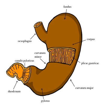
The stomach (ventriculus, gaster, stomachos) is a muscular sac located in the abdominal cavity under the left diaphragmatic vault. It extends upward under the rib cage into the regio hypochondriaca sinistra (the region of the abdomen below the diaphragm at the cartilage of the lower ribs), from there the stomach goes down to the right into the regio epigastrica (between the right and left rib arches).
It has the shape of a curved pouch with a left convex and a right concave edge (curvature):
- curvatura major (greater curvature)– curvature of the left edge, curving downwards to the left;
- curvatura minor (lesser curvature)– curvature of the right edge, concave upwards to the right;
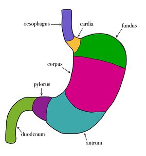
- cardia (comb) - the mouth of the oesophagus from above into the stomach at the curvature minor;
- pylorus (gatekeeper) - a narrowed place, adjacent to the stomach duodenum.
The three main parts of the stomach and their formations:
- fundus gastricus (ventriculi) – cranial, widest section, contains the food bubble;
- pars cardiaca – at the esophageal inlet, to the right of the fundus at the small curvature;
- ostium cardiacum (cardia) - the proper orifice of the esophagus, gastroesophageal junction;
- incisura cardiaca – the notch between the cardia and the fundus;
- corpus gastricum (ventriculi) – the body of the stomach;
- canalis gastricus – the body cavity of the stomach;
- incisura angularis – a break in the curvature of the curvature minor at the boundary of the fundus and pylorus (clearly visible on X-ray);
- pars pylorica –the distal section, the narrowest, passes into the duodenum;
- antrum pyloricum – beginning of pars pylorica at incisura angularis;
- canalis pyloricus – continuation of the antrum into its own gatekeeper, 2-3 cm long;
- pylorus (gatekeeper) - the place of transition of the stomach into the duodenum;
- ostium pyloricum – own (closable) opening of the stomach into the duodenum.
The empty or less filled stomach is flattened anteroposteriorly and is discernible on it:
- paries anterior – anterior wall (ventrocranially);
- paries posterior –posterior wall (dorsocaudally).
Functional sections recognized on a live stomach:
- pars digestoria – descending section, from cardia and fundus to incisura angularis, includes fundus and corpus, incisura major - incisure at curvature major, separates on the living fundus and corpus;
- pars egestoria – ascending section, from the incisura angularis right up to the pylorus (corresponding to the pars pylorica), 2 parts (separated by the sulcus intermedius) : the gastric sinus (corresponding to the antrum pyloricum) and the canalis pyloricus.
The peristalsis of the stomach takes place in four simultaneous waves, the largest being the contraction in the sulcus intermedius.
The shape of the stomach[edit | edit source]
- variable – individually, depending on the amount of filling and the position of the body;
- 2 typical shapes:
- hook-shaped (siphonic) stomach - "J" shape, more frequent, typical descending and ascending part, conspicuous incisura angularis;
- bull's horn-shaped stomach - oblique, smoothly tapering, gradual curvature.
Position and projection of the stomach[edit | edit source]
Skeleton projection[edit | edit source]
- Individually variable, ¾ stomach left of midline.
Position of the cardia[edit | edit source]
- Less variable;
- under the diaphragm to the left of the spine;
- lying down: projected in front of the left side of vertebra T10.
- standing: in front of the left side of vertebra Th12.
- fundus (fornix) with the stomach bubble from the cardia to the left, through the diaphragm urges on the heart and on the base of the left lung.
Position of the pylorus[edit | edit source]
- Standing: projected to the right side of the spine, most often to the level of the L2-L4 vertebrae.
- Lying down: approximately 2 vertebrae.
The lowest point on the curvature major[edit | edit source]
- Interface of vertebrae L3-L4, with bull's horn-shaped stomachs above, standing up to vertebra L5.
Projection on the anterior abdominal wall[edit | edit source]
Cardia[edit | edit source]
- Standing: projected on the left rib arch to the tip of the 7th rib, the upper edge of the fundus (fornix) extending to the level of the 5th-6th rib cartilages.
Curvatura major[edit | edit source]
- It crosses the left rib arch at the level of the tip of the attached cartilage of the 10th rib;
- caudally may reach close to the bicrystalline line.
Pylorus[edit | edit source]
- It is projected 2.5 cm to the right of the midline + 5 cm caudal to the centre of the right rib arch.
Relations of the stomach to its surroundings (syntopy)[edit | edit source]
The front surface of the stomach presses on:
- the lower surface of the liver (left lobe);
- left diaphragmatic vault;
- the anterior abdominal wall.
The posterior surface of the stomach presses on:
- diaphragm;
- left adrenal gland and left kidney;
- pancreas;
- spleen;
- mesocolon transversum.
The lower edge of the great curvature insists on:
Hinges on the stomach[edit | edit source]
- Duplications of the peritoneum (peritoneal double sheets);
- continue from the serous coating of the stomach from both curvatures as parts of the original anterior and posterior mesogastrium;
- connect the stomach to the environment;
- where the vessels of the stomach come to the curvatures.
Omentum minus (small vestibule) - part of the original anterior mesogastrium, peritoneal duplication extending from the curvature minor to the lower surface of the liver.
Bursa omentalis - space behind the omentum minus and stomach (between them and the peritoneal cover of the posterior abdominal wall), borders : liver (cranially), mesocolon transversum (caudally).
Omentum majus (large vestibule) - part of the original posterior mesogastrium, from the curvature major of the stomach → through the colon transversum (coalesces with it) → caudally in front of the intestinal villi (between them and the anterior abdominal wall) as a wide, freely hanging peritoneal lash, permeated by blood vessels or even adipose tissue, increasing the absorptive surface of the peritoneum.
- Lig. gastrocolicum – the section of the omentum majus between the stomach and its junction with the colon transversum;
- Lig. gastrolienale – part of the omentum majus, its continuation from the curvature major of the stomach to the left into the hilum of the spleen.
Stomach size[edit | edit source]
- variable;
- empty stomach: length 25 cm, width at fundus 4-5 cm, at pylorus 1.5 cm, weight 130 g, volume about 1 liter but can increase up to 3 liters.
Construction of the stomach wall[edit | edit source]
Structure in 4 layers (corresponds to the general structure of the digestive tube)
- mucosa - with many stomach glands - tunica mucosa
- submucosal ligament - tela submucosa
- muscle layer - tunica muscularis
- serous coating - tunica serosa
Mucosa of the stomach[edit | edit source]
- composed of epithelium (single-layered cylindrical), lamina propria mucosae (sparse collagenous connective tissue), lamina muscularis mucosae;
- orange-red on the living (pale, poisoned on the corpse);
- "junctional line" against the lighter esophageal mucosa;
- protective mucus on the surface;
- plicae gastricae (mucous membrane algae) - reticulate, with a predominance of longitudinal algae:
- longitudinal lashes - more conspicuous in both curvatures (highest in curvature minor);
- sulcus salivarius (Waldeyer's path) - the space between the longitudinal algae, the path from the cardia to the pylorus, liquid food flows through the empty stomach;
- articulation of the mucosal surface:
- areae gastricae – patches (2-6 mm) separated by inclusions;
- foveolae gastricae (stomach pits) - deep crypts lined with superficial epithelium of mucosa, stomach glands (2-7 pcs.);
- glandulae gastricae (gastric glands) - perpendicular to the surface of the mucosa, from the bottom of the pits in the form of tubular glands to the mucosal connective tissue to the lamina muscularis mucosae;
- glands of the stomach:
- glands in cardia - tubular, simple or poorly branched, cells produce thinner mucus - contains lysozyme;
- glands of the fundus and body of the stomach - simple, tubular, parts: base, proper gland, neck, isthmus, gastric pit, in the glands of the fundus 6 kinds of cells:
- mucus cells of isthmus - mucus neutral reaction;
- mucus cells of the neck - mucus acid reaction (glycosaminoglycans);
- undifferentiated cells - replace mucus cells of the isthmus and neck, surface cells in the gastric pit;
- main cells - lipases, pepsin (produced as pepsinogen - activated by acidity);
- cover cells - produce hydrochloric acid (HCl), an intrinsic factor;
- endocrine cells - serotonin;
- pyloric glands - tubular, often branched and coiled, shorter than the glands of the fundus and body of the stomach, produce mucus - similar to the mucus of the glands in cardia, contains lysozyme, G-cells - endocrine cells are interspersed among the exocrine cells of the gland, produce gastrin (release of acid in the glands of the fundus and body).
Submucosal connective tissue[edit | edit source]
- Sparse, allows shifting of the mucosa with changes in filling and movements of the stomach;
- contains networks of blood vessels and nerve plexus.
Musculature of the stomach[edit | edit source]
- Three layers:
- stratum circulare – thickest, thickening from the pars digestoria to the pars egestoria, strongest in the pylorus, where it forms: musculus sphincter pylori - 2 juxtaposed annular thickenings, connected to each other on the side of the curvature minor.
- stratum longitudinale – in pars digestoria reinforced along both curvatures in stripes: taenia curvaturae majoris et minoris, in pars egestoria forming a continuous mantle.
- fibrae obliquae – the innermost layer, the fascicles adjacent to the submucosa, run from the cardia obliquely to the curvature major.
- muscle function:
- maintains the shape of the stomach by its tension;
- peristolytic activity - tension of the walls and their adhesion to the contents of the stomach;
- peristaltic activity - produces annular contractions - like a wave they advance down the stomach from the cardia to the pylorus and move the contents of the stomach (they do not pass to the duodenum).
Serous coating of the stomach[edit | edit source]
- It consists of the peritoneum - smooth, shiny, passing from the curvature minor and major.
X-ray image of the stomach[edit | edit source]
- It shows on live the shape and position of the stomach and their variability;
- X-ray contrast slurry: BaSO4 - barium sulphate (image affected by volume and weight of the contrast material);
- also shows the peristalsis of the stomach wall.
- In X-ray imaging, the fundus is referred to as the fornix gastricus.
Blood vessels and nerves of the stomach[edit | edit source]
Arteries and veins[edit | edit source]
Curvatura minor
- arteria gastrica dextra et sinistra (← truncus coeliacus) (left and right gastric arteries)
- vena gastrica dextra et sinistra (→ vena portae) (left and right gastric veins)
curvatura major
- arteria gastroepiploica dextra et sinistra (left and right gastro-omental arteries)
- vena gastroepiploica dextra et sinistra (→ vena portae);
- arteriae gastricae breves – branches of arteria splenica.
Lymphatic vessels[edit | edit source]
- They start as a network in the mucosa (connected to a network in the submucosal connective tissue) – from there the lymph flows to another network in the muscular tissue and submucosal connective tissue;
- from a network of subserosal lymphatic collectors along the veins to individual lymph node groups: nodi lymphatici gastrici sinistri (left, at curvatura minor), nodi lymphatici gastrici dextri (right, at curvatura minor), nodi lymphatici gastroepiploici sinistri (left, at curvatura major), nodi lymphatici gastroepiploici dextri (right, at curvatura major);
- other groups of nodes: nodi lymphatici pancreaticolienales, nodi lymphatici pylorici;
- the lymph of all groups drains into the nodi lymphaticic coeliaci (at the truncus coeliacus).
Nerves[edit | edit source]
- Belongs to the autonomic nervous system, 2 groups:
Parasympathetic nerves[edit | edit source]
- They come by way of the right and left nervus vagus along the oesophagus, as the truncus vagalis anterior et posterior;
- of them to the anterior and posterior wall of the stomach: rami gastrici anteriores et posteriores - 1st neurons of innervation (with cells in the CNS), followed by 2nd (peripheral) neurons with cells in the gastric wall;
- their fibres form the plexus myentericus et submucosus;
- parasympathetic fibers increase muscle wall tension, peristalsis, and promote secretion of gastric glands;
- the vagal branches also contain sensory fibres (pressure, cold, heat).
Sympathetic nerve fibres[edit | edit source]
- They come to the stomach from the right and left sympathetic trunks by way of the splanchnic nerves and plexus coeliacus, and further along the vessels in the form of plexus;
- unconstantly come through the left nervus phrenicus;
- also sensory fibers (pain).
Links[edit | edit source]
Related articles[edit | edit source]
- Development of the stomach
- Tumours of the stomach
- Determination of gastric secretion, acidity
- Gastroduodenal ulcer disease
- Overview of gastrointestinal hormones
- Digestive system
- Stomach juice
- Filling, emptying and gastric motility
- Stomach as a histological specimen in czech:https://www.wikiskripta.eu/w/Portál:Sb%C3%ADrka_histologických_preparátů_(1._LF_UK)/Trávic%C3%AD_soustava
- Practice of histological preparations in czech:https://www.wikiskripta.eu/w/Procvičován%C3%AD:Trávic%C3%AD_systém
Used literature[edit | edit source]
- ČIHÁK, Radomír – GRIM, Miloš. Anatomie. 2. upr. a dopl edition. Grada Publishing, 2002. 470 pp. vol. 2. ISBN 80-247-0143-X.


