Plasma proteins
|
This article was marked by its author as Under construction, but the last edit is older than 30 days. If you want to edit this page, please try to contact its author first (you fill find him in the history). Watch the discussion as well. If the author will not continue in work, remove the template Last update: Saturday, 04 Dec 2021 at 7.58 pm. |
Proteins in blood serum or plasma are represented by many types of proteins produced by various different cells. The biosynthesis of the vast majority of plasma proteins takes place in liver, a smaller part is synthesized in other places, e.g. lymphocyte (immunoglobulins), enterocytes (e.g. apoprotein B-48) among others. Protein degradation takes place in hepatocyte and macrophages, where proteins are degraded predominantly after complex formation (e.g. antigen - antibody complex, hemoglobin - haptoglobin complex). Intracellularly, peptide bonds of proteins are hydrolyzed by proteases and peptidases to form amino acids. Another way to remove serum proteins is through the excretion of organs, especially the kidneys and gastrointestinal tract.
Total serum concentration of proteins is 65 to 85 grams per litre. Because plasma proteins are oncotically active, their physiological concentrations account for 3.33 to 3.52 kPa oncotic pressure (25 to 26.4 torr). The concentration of proteins in plasma is slightly higher then in serum because plasma contains coagulation factors.[1]
Functions of plasma proteins
Plasma proteins are necessary for a variety of blood/plasma functions:
- maintenance of oncotic pressure;
- transport of lipophilic compounds e.g. hormones (thyroid hormone bound to transtyretin; sex hormones), vitamins, lipids (bound to albumin), bilirubin bound to albumin, drugs);
- nutrition function;
- acidobasic buffer of blood;
- hemocoagulation a fibrinolysis;
- immunity
Overview of plasma proteins
|
Albumin
Albumin is the most common serum protein, it accounts for approximately 55 to 65% of total serum proteins (average blood concentration is 40 g/L[2]). Je syntetizován v játrech a jeho tvorba závisí na příjmu aminokyselin.
- Albumin is crutial for maintenance of the oncotic pressure of the plasma. Decreased albumin concentrations (hypoalbuminemia) below 20 g/L usually lead to oedemas.
- It acts as a carrier. It enables the transport of bilirubin, heme, steroid compounds, tyroxine, fatty acids, bile acids, metals, drugs, among others.
- Albumin is a protein reserve of the body and serves as a source of amino acids, especially essential amino acids for various tissues. During malnutrition, its concentration decreases, however, serum albumin levels are not a good indicator of early protein malnutrition because albumin has a long plasma half-life and large body stock. For this reason, albumin is a long-term marker of nutrition.
Synthesis of albumin
The synthesis of albumin has multiple steps. Preproalbumin, a precursor of albumin is synthesized by hepatocytes, but does not exit the cell cytoplasm. Subsequently, preproalbumin enters endoplasmatic reticulum, where it is transformed into proalbumin, the most abundant intracellular form of albumin. Then, proalbumin enters Golgi complex, where it is transformed into albumin and excreted our of the cell.[3]
Acute-phase protein
Acute-phase protein are proteins that are increased during acute inflammatory reaction of the organism, trauma, surgiceries, infarct of myocard, tumours, birth, increased stress of physical exercise. All abovementioned situations can induce the increase of acute-phase proteins, but the molecular response will vary based on the underlying cause (i.e. bacterial inflammatory reaction will increase different proteins than e.g. trauma).
Such mediators serve to ensure the overall response of the organism, mutual communication and regulation of ongoing events. Additionally, they also produces the "general symptoms" of inflammatory process (fever, muscle and joint pain). These substances are of clinical importance, the main synthesis arises as a result of known pathology, or when its concentration corresponds to the extent of tissue damage. Therefore, these substances may be used as markers for confirming/determining the origin of organism damage (whose symptoms may be relatively non-specific), determining the extent of the damage, and monitoring the course of the therapy.
Positive markers of acute phase response
This group encompases various proteins, whose blood (plasma) levels increase upon the beginning of the pathological process. They can be divided into following groups based on their effect on the organism (or their purpose in organism):
Immune complexes
- The purpose of some proteins of acute phase is to neutralize the xenobiotics (or microorganism) that caused the inflammation. Examples may be:
- C-reactive protein (CRP),
- complement, (all complement cascade proteins are increased, C3 and C4 have the higher physiological plasma concentrations, therefore, their increase if the most pronounced),
- tumor necrosis factor α (TNF-α), interleukin 1 (IL-1) and interleukin 6 (IL-6).
Proteins that prevent the colateral damage of the inflammatory response
- During inflammation, immune cells (such as phagocytes) release cytotoxic compounds that may damage not only the pathogen, but also the healthy tissues, which would cause undesirable side effects. To avoid this colateral damage, during inflammatory response, organism produces proteins that excessive inactivate proteolytic enzymes and reactive oxygen species (ROS) mitigating the damage to its own tissues. Such compounds include
- Protease ingibitors
- Compounds that decrease the synthesis or concentration of reactive oxygen species (ROS) - not only ROS scavengers, but also proteins that bind metal (mostly iron and copper) whose presence may worsen the inflammatory process. As a resuls, chelation of iron and copper may decrease the synthesis of reactive oxygen species (via Fenton reaction). Such compounds include
- Compounds, whose purpose is the transport of waste products away from the inflammation. Such compounds include
- hemoglobin
- hemopexin
- serum amyloid A (SAA).
- Coagulation factors and proteins inducing the tissue regeneration. Such compounds include
The purpose of some positive reactants, such as procalcitonin (PCT), remains unknown. Despite the unknown purpose, its increase of plasma concentration may be of a key importance when determining the nature of acute phase response. Therefore, even some compounds with unknown physiological function may be clinically useful.
The rate of increase of acute-phase proteins
The rate of increase of acute-phase proteins varies considerably. Therefore, for clinical purposes, we can divide acute-phase proteins into three groups: early, intermediate, and late based on the rate of increase of their plasma concentrations.
Časné proteiny akutní fáze
jsou bílkoviny s velmi krátkým biologickým poločasem. Změny jejich plazmatické koncentrace jsou patrné již za 6–10 hodin po začátku onemocnění. Vzestup vrcholí obvykle v průběhu druhého a třetího dne. Hlavními představiteli jsou především C-reaktivní protein (CRP) a sérový amyloid A (SAA). Nověji se v klinické praxi používá prokalcitonin (PCT).
- C-reaktivní protein
C-reaktivní protein (CRP) je jedním z nejdůležitějších reaktantů akutní fáze. Je to bílkovina, která hraje úlohu opsoninu. Své jméno získal díky tomu, že precipituje s tzv. C-polysacharidem pneumokoků.[4]
Plazmatická koncentrace CRP se zvyšuje již za 4 hodiny po navození reakce akutní fáze a v průběhu prvních dvou dnů jeho koncentrace vzroste i více než 100krát. Maximální koncentrace je dosaženo za 24–48 hodin, přibližně 24 hodin je i poločas CRP.[5]
Fyziologicky bývá plazmatická koncentrace do 8 mg/L.[6] Rychlý a vysoký vzestup CRP (typicky na hodnoty nad 60 mg/l) doprovází především akutní bakteriální infekce, méně obvykle také mykotické infekce. Virové infekce naproti tomu bývají charakterizovány relativně malým vzestupem CRP (zpravidla pod 40 mg/l).[7] Stanovení plazmatické koncentrace CRP proto napomáhá v rozhodnutí, zda zahájit léčbu antibiotiky.[4] Úspěšná antibiotická terapie se pak projeví rychlým poklesem CRP, naopak při neúspěšné léčbě přetrvává zvýšení.
Stanovením CRP lze odhalit riziko pooperační infekce. Třetí den po operaci má jeho koncentrace rychle klesat k normě. Přetrvávající zvýšení nebo jen částečný pokles, následovaný dalším zvýšením, naznačuje přítomnost infekce nebo jiné zánětlivé komplikace.
Mírný vzestup CRP provází i infarkt myokardu. Obecně lze také říci, že mírně elevované hladiny CRP (obvykle kolem 10 mg/l) patří mezi známky vysokého kardiovaskulárního rizika.[8] Sledování koncentrací CRP je užitečné i při monitorování autoimunitních onemocnění.[9]
Nevýhodou CRP je jeho nízká specifita. Na rozdíl od prokalcitoninu neinformuje o tíži orgánového postižení, nýbrž pouze o přítomnosti infektu. Vzájemně jsou se tyto dva markery nenahrazují, ale doplňují.
- Prokalcitonin
V posledních letech se ve výzkumu i v klinické praxi začíná jako reaktant akutní fáze využívat prokalcitonin (PCT). Tuto bílkovinu o 116 aminokyselinách a molekulové hmotnosti 13 000 fyziologicky tvoří C buňky štítné žlázy jako prekurzor hormonu kalcitoninu. Zejména při generalizovaných bakteriálních infekcích jej však začnou produkovat i další buňky, hlavně neuroendokrinní buňky plic a střeva, ale i buňky parenchymatózních orgánů a při sepsi prakticky všechny tkáně a typy buněk[10]. Koncentrace této bílkoviny pak v plazmě prudce stoupá. PCT uvolněný při sepsi není konvertován na kalcitonin.[11] Přesný fyziologický význam prokalcitoninu není zdaleka objasněn; předpokládá se, že se podílí na regulaci zánětu a má analgetické účinky. Poločas prokalcitoninu je 1 den a po imunitní stimulaci vzrůstá jeho sérová koncentrace již během 2–3 hodin asi dvacetinásobně. Zvýšení lze pozorovat jen při generalizovaných bakteriálních, mykotických a protozoárních infekcích, neobjevuje se u virových infekcí. S méně výrazným vzestupem se lze setkat u polytraumat, popálenin a po rozsáhlých břišních operacích.
Stanovení PCT
Provádí se vysoce citlivou imunoluminometrickou metodou, PCT-LIA (Luminescence ImmunoAssay). Jde o metodu se dvěma monoklonálními protilátkami, jednou proti C-terminální sekvenci prokalcitoninu (tzv. katakalcinu) a druhou proti centrální části prokalcitoninu (tj. proti kalcitoninu). Anti-katakalcinové protilátky jsou immobilizovány na povrchu zkumavky, anti-kalcitoninové protilátky jsou značeny luminescenční sondou (derivátem akridinu). Metoda vyžaduje luminometr, je k ní třeba 20 μl séra nebo plasmy.
Jako rychá metoda se používá imunochromatografický test na prokalcitonin (PCT-Q) v séru a plasmě. Je k němu třeba 200 μl séra nebo plasmy, výsledek je k dispozici za 30 minut. Tento test se doporučuje pro rychlou diagnostiku akutní pankreatitidy.
Orientační hodnoty PCT
Normální hodnoty (ng/ml) < 0,5; chronické zánětlivé procesy < 0,5–1; bakteriální infekce komplikovaná systémovou reakcí 2–10; SIRS 5–20; těžké bakteriální infekce – sepse, MODS 10–1000. Při protrahované sepsi přetrvává zvýšená hladina PCT, zatímco hladiny některých jiných cytokinů klesají.[11]
Neinfekční příčiny zvýšení PCT
Pooperační stav, mnohočetné trauma, úraz teplem, kardiogenní šok, u novorozenců prvních 48 h po porodu.[11]
Ze srovnání PCT, CRP, IL-6 a WBC vyplývá, že ukazatelem s nejvyšší senzitivitou a specificitou pro diferenciální diagnostiku infekční a neinfekční etiologie SIRS je prokalcitonin.[12]
Proteiny akutní fáze se střední dobou odpovědi
jsou proteiny, jejichž koncentrace se mění 12–36 hodin po začátku onemocnění a maxima je dosaženo ke konci prvního týdne. Patří k nim α1-kyselý glykoprotein (orosomukoid), α1-antitrypsin, haptoglobin a fibrinogen.
Pozdní proteiny akutní fáze
jsou zastoupeny složkami komplementu C3 a C4 a ceruloplazminem, u nichž se změny rozvíjí až po 48–72 hodinách po začátku onemocnění. Vzestup koncentrací je ve srovnání s oběma předchozími skupinami proteinů méně vyjádřen a vrcholu dosahují až po 6–7 dnech.
Negativní reaktanty akutní fáze
Negativní reaktanty akutní fáze jsou bílkoviny, jejichž hladiny se v průběhu akutní zátěže snižují. Hlavními zástupci jsou albumin, prealbumin a transferin. Pro sledování a hodnocení průběhu reakce na zátěž mají menší význam než pozitivní reaktanty. Často jsou však využívány jako kritérium syntézy bílkovin v játrech a jako ukazatelé malnutrice.
Imunoglobuliny
Protilátky (imunoglobuliny) jsou specifické globuliny krevní plazmy s elektroforetickou pohyblivostí β–γ. Vznikají v plazmatických buňkách jako humorální součást imunitní reakce na určitý antigen. Molekula imunoglobulinu má schopnost specificky vázat příslušný antigen, proti kterému se vytvořila. Po vazbě vzniká imunitní komplex. Kromě toho imunoglobuliny plní další funkce zahrnující např. vazbu komplementu, vazbu na neutrofilní leukocyty a makrofágy, aktivaci fagocytózy. Imunoglobuliny dělíme na 5 tříd – IgG, IgM, IgA, IgD a IgE. U třídy IgG byly popsány ještě podtřídy – IgG-1, IgG-2, IgG-3, IgG-4, jejichž funkce se liší. Rovněž třída IgA není jednotná, tvoří ji podtřídy IgA-1 a IgA-2. Základní struktura molekuly imunoglobulinu je tvořena dvěma stejnými těžkými řetězci (H-řetězce), označovaných podle jednotlivých tříd γ, μ, α, δ a ε, a dvěma lehkými řetězci (L-řetězce) κ a λ, které jsou pro jednotlivé třídy společné. Každá molekula imunoglobulinu obsahuje buď κ nebo λ řetězce. Při první infekci baktériemi či protozoy nastupuje v průběhu 2–3 dnů tvorba protilátek IgM, která je později během 5–7 dnů vystřídána tvorbou IgG se stejnou specifitou. Opakovaná infekce způsobí rychlé zvýšení hodnot IgG a malé zvýšení koncentrace IgM. Výrazné změny množství imunoglobulinů se projeví při elektroforetickém vyšetření jako:
- hypogamaglobulinemie (snížení vrcholu v oblasti γ);
- hypergamaglobulinemie;
- polyklonální (zvýšení vrcholu β-γ globulinu o široké bázi);
- monoklonální (úzký vrchol v oblasti β-γ globulinů).
Hypogamaglobulinemie
Hypogamaglobulinemie vzniká v důsledku zvýšených ztrát imunoglobulinů močí nebo střevem. Jinou závažnou příčinou je pokles tvorby imunoglobulinů, který může postihovat všechny nebo pouze jednotlivé třídy. Tyto defekty humorální imunity mohou být primární nebo sekundární a jsou příčinou závažných imunodeficitních stavů projevujících se opakovanými infekcemi s těžkým průběhem.
Metody stanovení bílkovin v séru
Základním vyšetřením proteinů v séru nebo plazmě je stanovení jejich souhrnné koncentrace – tzv. celkové bílkoviny. Při nálezu patologických hodnot a v dalších indikovaných případech následuje podrobnější vyšetření, které zahrnuje elektroforézu sérových bílkovin, imunofixaci a cílené stanovení koncentrace vybraných sérových proteinů.
Serum protein electrophoresis
Principle
Serum protein electrophoresis (SPEP) is a separation method based on a movement of charged particles within electric field. The compounds of interest need to be charged (i.e. they must either be ions, or ampholytes). Most proteins have ampholytic nature, therefore, their net charge can be positive or negative with variance of pH of the buffer during electrophoresis. Once a mixture of various charged molecules is exposed to a stationary electric field, individual ions will start moving towards either electrodes. The velocity of movement of ions depends on following factors:
- the charge of the molecule (positive ions move towards negative electrode, negative ions move towards positive electrode).
- magnitude of charge (the higher charge, the more the molecule is attracted to the electrode; if the net charge of a molecule is equal to zero, the molecule will not move at all)
- size of the compound or relative molar mass of the compound (molecules with higher molar mass will move slower that those with lower molar mass)
- voltage
Usually, a mixture of protein is separated in electrophoresis at pH 8.6 (using akaline buffer). Because izoelektric point of most serum proteins is near 5 to 6, at pH 8.6, all proteins are negatively charged, therefore, they will move towards the anode (positive electrode).
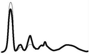
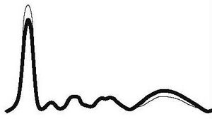
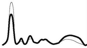
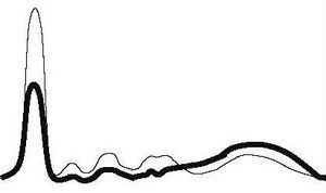
Blood serum electrophoresis usually separates proteins into 6 to 7 fractions: prealbumin (seen rarely), albumin, α1, α2, β1, β2, (sometimes poorly resolved, may be seen only as β fraction), γ fractions. With the exception of albumin and prealbumin fractions which contain only single protein each, these fractions consist of multiple proteins with similar electrophoretic mobilities.
Plasma protein concentration by electrophoretic fraction
| Fraction | Relative protein concentration (%) | Absolute protein concentration (g/L) |
|---|---|---|
| Albumin | 55 to 69 | 35 to 44 |
| α1 | 1.5 to 4 | 1 to 3 |
| α2 | 8 to 13 | 5 to 8 |
| β | 7 to 15 | 4 to 10 |
| γ | 9 to 18 | 5 to 12 |
Clinical use
Serum protein electrophoresis (SPEP) is used especially if we find a pathological result of the total protein, or if we need more detailed information about serum proteins. Especially valuable for the card:
- dysproteinemia - change in the concentration and qualitative composition of individual proteins in serum,
- paraproteinemia - characterized by the presence of monoclonal immunoglobulins.
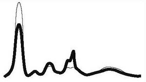
| Electrophoresis results | Comment | Alb | α1 | α2 | β | γ | Examples of common pathologies |
|---|---|---|---|---|---|---|---|
| Acute inflammation |
|
|
| ||||
| ↓ or N | ↑ | ↑ | N | ||||
| Chronic inflammation |
|
|
| ||||
| ↓ or N | N | N | N | ↑ | |||
| Chronic active inflammation |
|
|
| ||||
| ↓ | ↑ | ↑ | N | ↑ | |||
| Hepatic pathology |
|
|
| ||||
| ↓ | ↓ | ↓ | ↓ | ↑ | |||
| Nephrotic pathology |
|
|
| ||||
| ↓ | N | ↑ | ↑ | ↓ or N | |||
| Hypogamaglobulinemia |
|

|
| ||||
| N | N | N | N | ↓ | |||
| Monoclonal gamapathy |
|

|
| ||||
| ↓ | ↓ | ↓ | ↑ | ↑ | |||
Links
Reference
- ↑ BURTIS, Carl A a Edward R ASHWOOD. Tietz textbook of clinical chemistry. 2. vydání. Philadelphia : Saunders, 1994. 2326 s. ISBN 0-7216-4472-4.
- ↑ ŠVÍGLEROVÁ, Jitka. Albumin [online]. Poslední revize 2009-02-18, [cit. 2010-10]. <https://web.archive.org/web/20160416224413/http://wiki.lfp-studium.cz/index.php/Albumin>.
- ↑ RACEK, Jaroslav, et al. Klinická biochemie. 2. vydání. Praha : Galén, 2006. 329 s. s. 71. ISBN 80-7262-324-9.
- ↑ Jump up to: a b ZIMA, Tomáš, et al. Laboratorní diagnostika. 2. vydání. Praha : Galén a Karolinum, 2007. 906 s. ISBN 978-80-246-1423-6.
- ↑ ZIMA, Tomáš, et al. Normální hodnoty [online]. Velký lékařský slovník online, [cit. 2020-02-13]. <http://lekarske.slovniky.cz/normalni-hodnoty>.
- ↑ KESSLER, Siegfried. Laboratorní dagnostika. 1. vydání. Praha : Scientia medica, 1993. 252 s. Memorix; s. 52. ISBN 80-85526-12-3.
- ↑ KESSLER, Siegfried. Laboratorní dagnostika. 1. vydání. Praha : Scientia medica, 1993. 252 s. Memorix; s. 52. ISBN 80-85526-12-3.
- ↑ GREGOR, Pavel a Petr WIDIMSKÝ, et al. Kardiologie. 2. vydání. Praha : Galén, 1999. 595 s. s. 168. ISBN 80-7262-021-5.
- ↑ KLENER, Pavel, et al. Vnitřní lékařství. 3. vydání. Praha : Galén a Karolinum, 2006. 1158 s. ISBN 80-7262-430-X.
- ↑ LIU, H. H., J. B. GUO a Y. GENG. Procalcitonin: present and future. Irish Journal of Medical Science (1971 -). 2015, roč. 3, vol. 184, s. 597-605, ISSN 0021-1265. DOI: 10.1007/s11845-015-1327-0
- ↑ Jump up to: a b c ÚKBLD 1. LF a VFN Praha. Prokalcitonin : vývoj názorů na interpretaci [online]. ©2009. [cit. 2011-06-30]. <http://www.cskb.cz/res/file/akce/sjezdy/2009-Pha/ppt/B1/Kazda.pdf>.
- ↑ STRICKLAND, RD, ML FREEMAN a FT GURULE. Copper binding by proteins in alkaline solution. Analytical chemistry [online]. 1961, vol. 33, no. 4, s. 545-552, dostupné také z <https://pubs.acs.org/action/cookieAbsent>. ISSN 0003-2700. DOI: 10.1021/ac60172a019.


