Porphyria


|
This article was marked by its author as Under construction, but the last edit is older than 30 days. If you want to edit this page, please try to contact its author first (you fill find him in the history). Watch the discussion as well. If the author will not continue in work, remove the template Last update: Thursday, 06 Jan 2022 at 7.27 pm. |
Porphyrias are a group of metabolic diseases with a disorder of heme synthesis, characterized by the accumulation or increased excretion of porphyrins. Porphyrias can be divided into erythropoietic, hepatic and erythrohepatic. The most common of these diseases is porphyria cutanea tarda.[1]
Repetitorium of biochemistry - porphyrins
Porphyrins are cyclic compounds formed by four pyrrole rings joined by methine (= CH-) and in porphyrinogens by methylene bridges (-CH2-). Subsequently, they form complexes with metal ions (eg Fe in hemoglobin, myoglobin, cytochromes, catalase; Co in vitamin B12; Mg in chlorophyll).
They are colored in various shades of red (unlike colorless porphyrinogens). The coloration is given by the system of conjugated double bonds in their molecule, which absorb visible light. They fluoresce red in UV light. Due to the indispensability for life, it must be de novo synthesized in the body (ingested in food decomposes and serves as a source of nutrients).
Characteristics of porphyrias
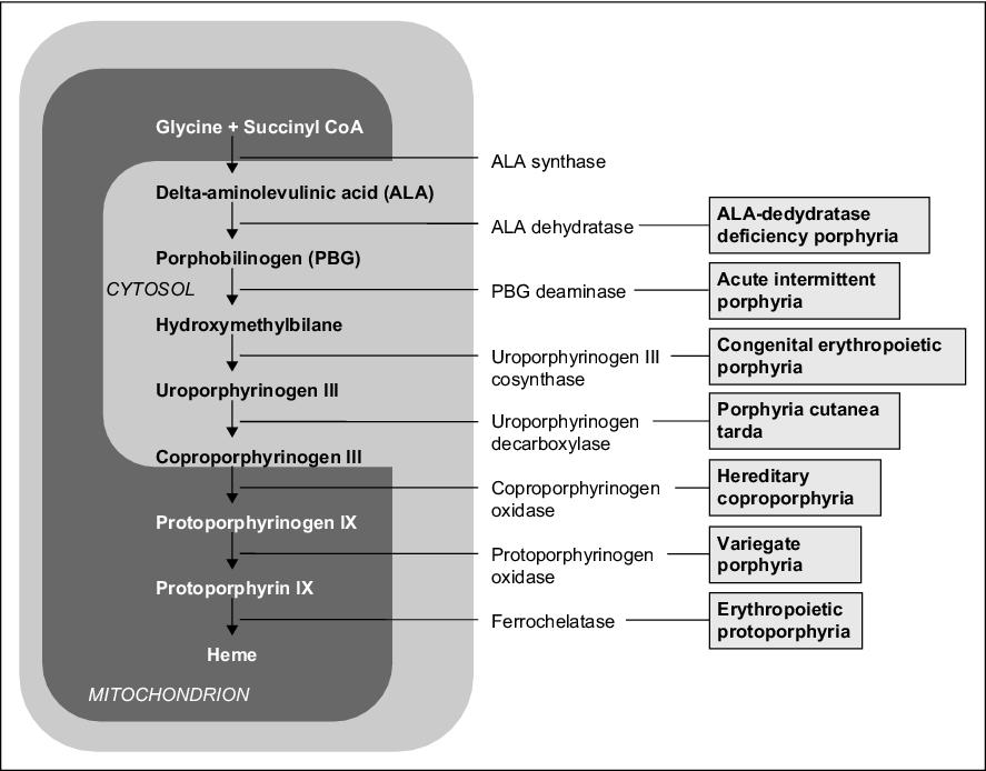
Mutations in the genes that control the synthesis of enzymes involved in heme synthesis lack the metabolic product behind the block (heme) or accumulate the metabolite in front of it.
In the case of an enzyme defect in the early phase of biosynthesis (before the formation of porphyrinogens), the starting substances ALA - δ-aminolevulinic acid (5-aminolevulinic acid) and PBG - porphobilinogen accumulate in body fluids, which have a toxic effect (interfere with synaptic functions) on the PNS and CNS , which creates typical symptoms - neuropathy (muscle weakness), abdominal pain, increased sympathetic activity and neuropsychic problems (restlessness, hysteria, psychotic states).
Defects in higher stages of synthesis lead to the accumulation of porphyrinogens, whose oxidation products (corresponding porphyrins) cause photosensitivity (hyperreaction to visible light in the 400 nm region). When porphyrins are exposed to light of this wavelength, they are excited, reacting with molecular oxygen to form oxygen radicals, which damage cellular organelles, including lysosomes → lysosomal enzymes are released, which damage light-exposed skin (erythema, blisters, scarring). Symptoms of the disease also include redness or browning of the teeth, urine and stool, or receding gums.
Individual methods of elimination are important in the diagnosis of porphyrias. ALA, PBG, uroporphyrin are soluble in water → excreted in the urine. Protoporphyrin is insoluble in water → excreted in the bile. Coproporphyrin is found in both bile and urine, and its urinary excretion increases with liver damage.
Manifestation is often only in adulthood after a certain evoking moment (drugs, sun exposure, stress, hormonal effects). The administration of some drugs (barbiturates, anesthetics) as well as alcohol increases the activity of ALA-synthase, which leads to increased production of porphyrins, and thus worsening of symptoms in patients with porphyrias (possibly acute attack). Acute porphyrias are more often affected by women and chronic men.
In acute therapy, glucose suppresses the induction of ALA synthase, the effect being potentiated by the application of heme derivatives such as heme-albumin or heme-arginate or hematin. Heme inhibits negative feedback ALA-synthase.[2]
It is divided according to:
- most affected organs producing porphyrins:
- hepatic - AIP, PCT, HCP, VP, ADP;
- erythropoietic - CEP;
- erythrohepatic - EPP;
- manifestations:
- skin - PCT, EPP, CEP; VP and HCP also cause skin manifestations;
- liver - AIP, ADP, VP, HCP;
- course:
- acute - AIP, VP, HCP, ADP;
- chronic - PCT, EPP, CEP;
| Porphyria type | Affected enzyme | The main symptoms | Laboratory finding |
| Acute intermittent (hepatic) | uroporphyrinogen I cosynthetase | abdominal pain, neuropsychiatrist. symptoms, not photosensitivity | U-PBG ↑,
U-uroporfyrin ↑ |
| Congenital erythropoietic | uroporphyrinogen III cosynthetase | photosensitivity | U-uroporfyrin ↑,
U-PBG ↓ |
| Porphyria cutanea tarda (skin) | uroporphyrinogen decarboxylase | photosensitivity | U-uroporfyrin ↑ |
| Porphyria variegata (hepatic) | protoporphyrinogen oxidase | photosensitivity, abdominal pain, neuropsychiatric symptoms | U-PBG ↑,
F-protoporfyrin ↑ |
| Protoporhyria (erythrohepatic) | ferrochelatase | photosensitivity | F-protoporfyrin ↑,
Ery-protoporfyrin ↑ |
| Hereditary coproporphyria (hepatic) | coproporphyrinogen oxidase | photosensitivity, abdominal pain, neuropsychiatric symptoms | U-PBG ↑,
U-uroporfyrin ↑ |
Porphyria skin
Porphyria cutanea tarda (PCT)
It is an AD hereditary defect of uroporphyrinogen decarboxylase, occurring in a ratio of 1:25 000 (the most common form), especially in middle-aged men. Porphyrins are produced in excess in the liver, they accumulate there, they are transmitted through the bloodstream to the skin, where they cause photosensitivity, which is a typical symptom. After exposing the skin to sunlight, large blisters filled with fluid appear, which heal very slowly with the formation of scars and mildew (dotted whitish deposits). The skin is hyperpigmented, later atrophic, easily vulnerable. Hypertrichosis occurs in the temples and around the eyes. The clinical manifestation is associated with liver damage caused by alcohol, polyhalogenated hydrocarbons (hexachlorobenzene, dioxin), estrogen treatment, hepatomas, hemochromatosis or hepatitis. Untreated can lead to liver cancer. There is also a non-hereditary form (sporadic, so-called PCT type 1). In the urine we find uroporphyrin, high levels of iron, in 50% of cases high levels of liver enzymes.
Treatment: repeated venipunctures (300-500 ml at 2-4 week intervals) depriving the body of excess porphyrins and iron + administration of antimalarial chloroquine (125-250 mg daily), which causes slow leaching of porphyrins, then protection from the sun (clothing, special creams) and a liver diet.
Congenital erythropoietic porphyria (CEP, Günther's disease)
This is an AR hereditary defect of uroporphyrinogen-III-synthase (UROS) leading to increased production of porphyrins in the bone marrow, which accumulate in the body, especially in erythrocytes. The incidence is 1: 2-3 million. This disease usually manifests itself in childhood. Manifestations of the disease vary - these include dark red urine (due to the presence of uroporphyrin and coproporphyrin), skin sensitivity (blistering, scarring) and darkening, eye sensitivity, eyelash loss, anemia, splenomegaly, red teeth, excessive hair growth ( especially on the hands and face).
Treatment: bone marrow transplantation, sun protection, blood transfusion, splenectomy.
.jpg)
Protoporphyria (EPP)
This is an AD hereditary defect of ferrochelatase, which results in the accumulation of protoporphyrin in the liver, bone marrow and skin. The most common symptoms are redness, itching and swelling of the skin even after a short (several minutes) exposure of the skin to sunlight. The symptoms disappear after hours to days, with repeated exposure there is scarring of the skin and other variable skin manifestations. The disease usually manifests itself in childhood. In a few percent of cases, liver damage occurs.
Treatment: alleviation of symptoms with beta-carotene, antihistamines, melanotan, phototherapy; prevention is the use of protective clothing and special creams. In contrast to acute hepatic porphyrias EPP.
Liver porphyria
Acute intermittent porphyria (AIP)
The basis for the emergence is an AD hereditary defect in hydroxymethylbilane synthase (by other names porphobilinogen deaminase, PBGD or uroporphyrinogen-I-synthase) leading to the accumulation of heme precursors in the liver. It manifests an acute attack after exposure to certain chemicals (steroids, drugs, alcohol), starvation, infection or stress; mostly in puberty. The main symptoms are abdominal pain (imitating NPB), constipation, vomiting, hypertension and mental problems (hysteria), headaches, paresis and plegia. There is an increased level of ALA and PBG in the urine. The blood is dominated by hyponatremia, hypokalemia with abnormalities in the metabolism of sugars and fats. The diagnosis is confirmed by decreased PBGD activity in erythrocytes.
Treatment: in the acute phase of infusion with glucose (inhibits ALA-synthase) and hematin; prevention of another attack is the avoidance of the inducing substance (ban on the use of certain drugs or alcohol).
Porphyria from 5-aminolevulate dehydration deficiency (ADP, Doss porphyria)
It is caused by an AR hereditary deficit of 5-aminolevulate dehydration.
Symptoms include abdominal pain and neuropsychiatric problems. ALA and coproporphyrin are present in the urine.
Hereditary coproporphyria (HCP)
It is caused by AD hereditary defect of coproporphyrinogen oxidase.
Symptoms include neuropsychiatric problems, photosensitivity, rarely abdominal pain, but completely asymptomatic forms are also common. In the acute stage, there are increased levels of ALA, PBG, coproporphyrin in the urine (this is also detectable in the stool).
Porphyria variegata (VP)
The etiology of AD is an inherited protoporphyrinogen oxidase defect
Symptoms include abdominal pain, neuropsychiatric problems and, in some patients, skin symptoms (photosensitivity). High levels of ALA, PBG, coproporphyrin in urine; increased excretion of protoporphyrin and coproporphyrin in faeces.
Diagnosis
By building up porphyrins in body fluids, urine and faeces (specific fluorescence).
Note: Porphyrinuria and ALA secretion may also be a symptom of lead poisoning (inhibits ALA dehydratase and ferrochelatase), resulting in anemia and ATP deficiency.
Porphyria treatment
Patients should pay attention to a proper lifestyle - enough vitamins; Avoid alcohol, garlic (contains enzymes that worsen the symptoms of porphyria), sunlight and UV radiation. Blood transfusions and heme injections are used for symptomatic treatment.
Links
Related articles
External links
Source
MASOPUST, Jaroslav a Richard PRŮŠA. Patobiochemie metabolických drah. 2. vydání. Univerzita Karlova, 2004. 208 s. s. 118–119.
Reference
Použitá literatura
- MURRAY, Robert, Daryl GRANNER a Peter MAYES, et al. Harperova biochemie. 4. české vydání. Jinočany : Nakladatelství H+H, 2002. 872 s. s. 354-360. ISBN 80-7319-013-3.
- LEDVINA, Miroslav, Alena STOKLASOVÁ a Jaroslav CERMAN. Biochemie pro studující medicíny : II.díl. 2. vydání. Praha : Nakladatelství Karolinum, 2009. s. 336-341. ISBN 978-80-246-1415-1.
- MASOPUST, Jaroslav a Richard PRŮŠA. Patobiochemie metabolických drah. 1. vydání. Praha : Univerzita Karlova, 2. lékařská fakulta, 1999. 182 s. s. 104-110. ISBN 80-238-4589-6.
- KALOUSOVÁ, Marta, et al. Patobiochemie ve schématech. 1. vydání. Praha : Grada Publishing a.s, 2006. 264 s. s. 51-57. ISBN 80-247-1522-8.
