Candidosis


Candidosis (candidiasis) is a common infection caused by a yeast of the genus Candida. It mainly affects intertriginous localizations of the skin and mucous membranes.[1]
Candida spp. belongs to the nosoparasitic fungi that live as saprophytes on the skin, oral cavity, vagina or intestine of humans. Under certain conditions, they multiply and then cause the disease. These conditions include: heat, moisture, maceration, skin surface disorders, pH changes (especially alkalization), obesity, pregnancy, diabetes mellitus, cachectizing diseases, antibiotics, gluco-corticoids or immunodeficiencies.
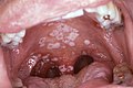
Soor (oral candidiasis)
White coatings on the lining of the tongue, cheeks or palate, may spread to the pharynx, and exceptionally to the stomach. The original white color changes through yellow to brown. The coatings can be wiped off at first, later they adhere firmly. Mucosa around is deep red, it usually spreads to the corners of the mouth.
Occurrence in toddlers, in immunodeficient, in patients who are treated with broad-spectrum antibiotics.
Anguli infectiosi
Most often caused by Candida. Interacting factors: deep bite, diabetes mellitus, ariboflavinosis, GIT disorders and more. We distinguish differentially diagnostically: mouth corners, syfilis, herpes simplex.
Therapy: nystatin, borax-glycerin superficially, amphotericin B.
Vulvovaginitis candidosa
Occurs in older women. Reddening of the mucosa, swelling of the vulva, severe itching, whitish coatings and discharge, rectal infections.
Balanitis candidosa
More common in diabetics. Red, eroded, sharply demarcated itchy deposit, whitish coatings.
Candidosis intertriginosa
Most often in the genitocural grooves, in the lashes and under the breasts. Manifestations of itching and burning, areas reddish, sharply demarcated, in the vicinity of the sowing of itching blisters.
Erosio interdigitalis candidosa (blastomycetica)
Professional diseases of cooks, bakers and confectioners. Mainly between the 3rd and 4th finger on the hand. Red, itchy erosion or ragade, whitish, macerated epidermis around. Maceration and sugar contribute to it.
Paronychia candidosa
Also professional disease. Chronic inflammatory swelling and redness of eponychium (nail fold), from which a drop of whitish exudate can be squeezed out.

Oral candidiasis (soor)

Candidiasis of the tongue
Oral candidiasis (soor)

Candidate paronychium

Candida albicans


Video
Links
Related articles
External links
Source
- BENEŠ, Jiří. Studijní materiály [online]. ©2007. [cit. 2015-03-24]. <http://www.jirben2.chytrak.cz/materialy/dermatovenerologie.doc>.
References
- ↑ ŠTORK, Jiří. Dermatovenerologie. 2. edition. Galén, 2013. 502 pp. ISBN 978-80-7262-898-8.
Bibliography
- ŠTORK, Jiří. Dermatovenerologie. 2. edition. Galén, 2013. 502 pp. ISBN 978-80-7262-898-8.
- LOBOVSKÁ, Alena. Infekční nemoci. 1. edition. Karolinum, 2001. 263 pp. ISBN 80-246-0116-8.
- HAVLÍK, Jiří. Infektologie. 2. edition. Praha : Avicenum, 1990. 393 pp. ISBN 80-201-0062-8.
