Skin efflorescence
Efflorescence is a skin manifestation of the disease. Individual efflorescences can merge into disease centers or areas. A collection of efflorescences is called an exanthema. If the exanthema is on the mucous membrane, it is called an enanthema. Primary efflorescence is the initial manifestation of the disease. Secondary will arise from the development of primary efflorescence or as a secondary manifestation on deposits or surfaces.
For efflorescence we evaluate:
- localization,
- size,
- face,
- color,
- Surface,
- border,
- Surroundings.
Primary efflorescence[edit | edit source]
A macula (spot) is a small area change in color mainly caused by a change in blood supply, the amount of blood vessels or pigment.
- freckles, vitiligo, some drug rashes
A papule is a small skin prominence up to 1 cm.
- psoriasis, warts
- papulopustula − a bud containing a cavity filled with pus (acne)
- papulovesicle − a bud with a cavity with clear contents
A tuber (bump) is a papule of larger dimensions (over 1 cm).
- in furuncle, spinalioma, lipoma
Pomphus (nettle bud, urtica) is a bud caused by edema of the corium, lasts a short time, usually itches.
- urticaria, insect sting (mosquito)
A vesicle is a cavity filled with clear contents.
- subcorneal − impetigo
- intraepidermal − pemphigus vulgaris , eczema , herpes simplex
- subepidermal − bullous pemphigoid , dermatitis herpetiformis (Duhring)
The bulla is larger, it arises, for example, in Lyell's syndrome, the bullous form of impetigo
A pustule is a cavity filled with pus; but more often it arises as a secondary clouding of the contents of the vesicle
- primary − folliculitis, psoriasis pustulosa
A nodule is a circumscribed round formation that arises from changes in the corium or subcutaneous tissue. It may be palpable beneath the surface or may be raised.
- E.g.: erythema nodosum, lipoma.
Secondary efflorescence[edit | edit source]
Squama (scale) is a thin plate of exfoliating horn.
- pityriaziform - bran-like scales (pityriasis versicolor)
- lamellar - lamellar (chronic eczema)
- exfoliative − striated ( sunburn )
A crust (scab) is formed by the drying of tissue fluid (yellow color), pus (yellow-green color) or blood (brown to black color).
- crustosquama − a scale penetrated by liquid and dries up
- rupia − terraced layered
An eschar is caused by the death of the skin, the dead tissue is initially whitish, later gray-brown to black, bordered by demarcation inflammation, leaves an ulcer, heals with a scar.
- eg: burns, burns, trophic circulatory disorders
Ragada (fissure) is a slit-like violation of the continuity of the skin, it is caused by a rupture of the epidermis only ; if it goes under the epidermis, it bleeds − fissura (crack).
- with impaired skin elasticity (edema, infiltrate, hyperkeratosis...), in places of mechanical stress
Erosion (abrasion) - a superficial defect affecting only the epidermis or mucous membrane, when the stratum spinosum is exposed, it oozes, breaking the papillae will cause slight bleeding.
Excoriation is caused by mechanical scratching (during pruritus).
Ulcus (ulcer) is a deep defect of the skin, it always affects the corium or deeper, we describe the size and shape, the base (clean, coated; smooth, bumpy,...)
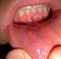
Aphtha is an efflorescence occurring only in the oral cavity, it is an erosion covered by fibrin, bordered by a red border.
Other terms when describing skin changes[edit | edit source]
Erythema - red discoloration of the skin.
Oedema (edema) – diffuse swelling of the skin.
Madidatio (wetting) – sprinkling of tissue fluid .
Cicatrix (scar) − is created by the transformation of granulation tissue (loss of fibroblasts and capillaries), fresh is pink, old is whitish. The scar can be atrophic, hypertrophic, keloid.
Lichenification is a thickening of the skin.
Links[edit | edit source]
[edit | edit source]
Source[edit | edit source]
- BENEŠ,. Study materials [online]. ©2007. [cit. 9.12.2010]. <http://jirben2.chytrak.cz>.
Literature[edit | edit source]
- ŠTORK, Jiří. Dermatovenerology. 1. edition. Prague : Galén, Karolinum, 2008. ISBN 978-80-7262-371-6.
- JIRÁSKOVÁ, Milena. Dermatovenereology for dentists. 1. edition. Praha : Professional Publishing, 2001. ISBN 80-86419-07-X.


