Eye (histology)
From WikiLectures
- a complex, highly specialized organ. It enables accurate analysis of object form, light intensity and colors.
- stored in orbit
- composed of:
- The eyeball (bulbus oculi) , in which we also find the so-called refractive structures of the eye
- Accessory structures of the eye (organa oculi accessoria)
The eyeball (bulbus oculi)[edit | edit source]
It consists of 3 layers:
- tunica fibrosa, which consists of:
- cornea
- sclera
- tunica vasculosa, consisting of 3 other parts:
- choroid
- ciliary body
- iris
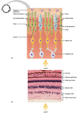
- tunica nervosa, formed by the retina, which consists of 2 parts:
- pars caeca retinae
- pars optica retinae
Tunica fibrosa[edit | edit source]
- Cornea
- thicker than the sclera
- colorless
- transparent
- avascular
- composed of 5 layers:
- Anterior corneal epithelium - multi-layered squamous non-keratinizing (5-6 cell layers)
- Bowmann's membrane – formed by collagen fibers; is acellular
- Substantia propria corneae – 50 to 60 layers of bundles of parallel collagen fibrils that cross at approximately right angles
- Descemet's membrane - has the character of a basal lamina
- Posterior corneal epithelium – single-layer flat
- corneal epithelia are able to transport ions across the cell membrane
- the regular arrangement of collagen fibrils ensures the transparency of the cornea
Tunica vasculosa[edit | edit source]
- highly vascularized layer
- there is also a sparse collagen fiber rich in fibroblasts and melanocytes
- Choroid (choroidea)
- has 4 layers:
- Lamina suprachoroidea – a layer of thin collagenous tissue
- Zona vasculosa
- Lamina choriocapillaris – anastomosing network of capillaries
- Lamina vitrea (Bruch's membrane)
- Corpus ciliare
- Orbiculus ciliaris – muscle ciliaris
- Corona ciliaris – sparse collagen tissue
- Iris
- consists of 4 layers:
- Anterior iris epithelium
- Front boundary layer
- Stroma iridis
- Pars iridica retinae
Tunica nervosa[edit | edit source]
- Pars ceaca retinae
- pars ciliaris retinae
- pars iridica retinae
- Pars optica retinae
- has 10 layers (ordered from outermost to innermost)
Refractive structures of the eye[edit | edit source]
- consists of:
- lens (lens crystallina)
- humor aquaeus
- corpus vitreum (vitreous) – transparent gel, 99% water, it contains hyalocytes.
Lens (lens crystallina)[edit | edit source]
- transparent
- biconvex
- consists of 3 parts:
- capsula lentis – covers the entire lens, contains type IV collagen
- anterior epithelium of the lens – located only on the anterior surface
- lens fibers – hexagonal prism shape
Accessory structures of the eye[edit | edit source]
- conjunctiva
- eyelids (palpebrae)
- lacrimal apparatus
- oculomotor muscles
Conjunctiva[edit | edit source]
- thin, transparent
- lines the conjunctival sac
- lined with multilayered cylindrical epithelium
Eyelids (palpebrae)[edit | edit source]
- moving bodies
- they are covered by skin on the outside, conjunctiva on the inside
- the eyelashes depart from the free edge
- the basic supporting structure is the tarsus, which is made up of dense collagenous tissue
- striated muscle is found here
- contains 3 types of glands:
- Meibomian
- long, branched, alveolar, sebaceous
- opens into the conjunctival sac
- Zeiss
- small, branched, alveolar, sebaceous
- opens into the eyelash follicle
- Moll's
- modified, simple, tubular, coiled, apocrine
- opens into the eyelash follicle
- Meibomian
Lacrimal apparatus[edit | edit source]
- consists of the lacrimal gland and the duct system
- the function of the lacrimal gland is to moisten the surface of the eye
- glandula lacrimalis – compound tuboalveolar gland, its serous compartment consists of cylindrical cells of the serous type, and its ducts merge into
- ductuli lacrimales, which are lined with a single-layer cubic epithelium and open into
- conjunctival sac, from which tears are drained using
- lacrimal canals (canaliculi lacrimales), they are lined with multi-layered squamous epithelium and merge into one canal and open into
- saccus lacrimalis, which continues as
- ductus nasolacrimalis and opens into the meatus nasi inferior
- saccus lacrimalis and ductus nasolacrimalis are lined with multi-rowed cylindrical epithelium, which is found in the greater part of the lining of the respiratory system
Links[edit | edit source]
Related articles[edit | edit source]
- Eye (biophysics)
- Eye (biophysics)/Principle of vision
- Eye (biophysics)/Disorders of the eye
- Oculomotor muscles
Sources[edit | edit source]
- MESCHER, Anthony L. Junqueira's Basic Histology. 12. edition. McGraw-Hill Education - Europe, 2009. pp. 480. ISBN 9780071630207.
- KONRÁDOVÁ, Václava – UHLÍK, Jiří – VAJNER, Luděk. Funkční histologie. 2. edition. H & H, 2000. pp. 291. ISBN 80-86022-80-3.
- PAULSEN, Douglas F. Histologie a buněčná biologie : Opakování a příprava ke zkouškám. 1. edition. H & H, 2004. pp. 433. ISBN 80-7319-024-9.


