Hypercalcemia
Hypercalcemia refers to a serum calcium level higher than 2.8 mmol / l with ionized calcium being higher than 1.4 mmol / l.
Hypercalcemic crisis is a life-threatening condition with a calcemia higher than 4 mmol / l.
Hypercalcemia:
Etiology[edit | edit source]
YouTube video on hypercalcemia
- Primary and tertiary hyperparathyroidism
- malignant tumors : lymphomas, leukemia, osteolytic metastases, paraneoplastic syndrome
- other endocrinopathies : hyperthyroidism, Addison's disease, pheochromocytoma, acromegaly,
- granulomatous diseases : sarcoidosis, tuberculosis, histoplasmosis, candidiasis, Wegener's granulomatosis, subcutaneous fat necrosis,
- pharmaceuticals : thiazides, milk-alkali syndrome, milk-alkali syndrome, vitamin D, A intoxication, acetylsalicylic acid,
- familial hypocalciuric hypercalcaemia,
- Williams-Beuren syndrome,
- Paget's disease,
- immobilizers,
- hypophosphatemia,
- transplant rejection.
Clinical picture[edit | edit source]
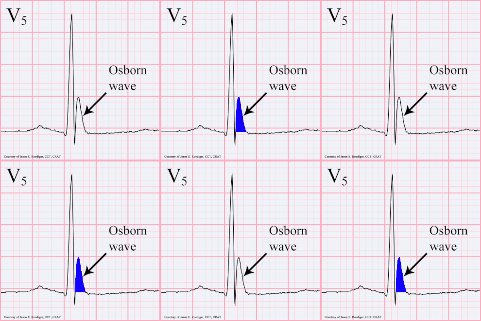
Hypercalcemia is manifested by a wide variety of GIT symptoms- nausea, vomiting, metallic taste in the mouth, gastroduodenal ulcer disease, persistent constipation, pancreatitis. In terms of kidney disease, we find polyuria, polydipsia, nocturia, and sometimes even symptoms of chronic or acute renal insufficiency. Hypercalcemia is also manifested in the CNS: we find headaches and disorders of consciousness. Neuromuscular symptoms include weakness and limb muscle pain. Cardiovascular symptoms include hypertension, arrhythmias, and ECG changes (shortening of the QT interval). Changes on the ECG confirm the severity of hypercalcemia, which in extreme cases can also cause cardiac arrest in systole - we speak of the so-called " chemical death " at a calcium level > 4 mmol / l. Other symptoms of hypercalcemia include pallor and, as seen on a X-ray image of bones, a significant zone of temporary calcification, osteoporosis, metastatic calcifications.
Diagnosis[edit | edit source]
We determine the ionogram - ions: Ca, P, Na, Cl, Mg. The physiological level of serum phosphorus is 0.65–1.6 mmol and for infants it is 1.3–2.3 mmol / l. Decreased phosphorus or values at the lower limit of normal are found in hyperparathyroidism and, conversely, increased phosphorus or at the upper limit of normal is found in hypervitaminosis D or thyrotoxicosis . A simple test is the S-Ca / S-P ratio. If this index is> 27% , it indicates hyperparathyroidism, if it is lower, it indicates hypercalcemia from other causes. The next step is to determine ALP and its specific isoenzymes. Elevation of ALP in hypercalcemia indicates increased bone resorption (hyperparathyroidism, malignancy). Hyperchloremic metabolic acidosis indicates hyperparathyroidism. Other detailed examinations include determination of urinary concentrations (U-Ca, UP, U-cAMP, U-citric acid), bone densitometry, and determination of immunoreactive PTH. Imaging methods include wrist and forearm X-ray, S + P X-ray, parathyroid ultrasound, abdominal ultrasound, abdominal CT. The Ca / creatinine index (so-called Nordin index) using morning urine is helpful: in healthy children it is up to 0.6, in infants up to 1.0. During hypercalciuria the values are higher, while in hypocalciuria the value is <0.2. If sarcoidosis is suspected, we can also use the hydrocortisone test : we administer 100 mg of hydrocortisone daily for 7 days. If there is a decrease in the hypercalcemia, then the underlying disease is most likely sarcoidosis. Malignancy screening should also be performed if hypercalcemia is of unclear origin.
Therapy[edit | edit source]
In the treatment of acute hypercalcemia, we primarily administer saline 20 ml / kg iv within 60 minutes. The goal is hyperhydration, where we calculate the physiological daily fluid requirement as twice the norm. Following the bolus of saline, we administer the diuretic furosemide 1-2 mg / kg iv and we repeat this after 2-3 hours. In any case, we must carefully monitor fluid balances and consider the risk of hypokalemia during drastic diuretic therapy. It follows from the above that in the next procedure, we supplement the circulating volume with diuresis losses and compensate for potassium losses.
A reduction in hypercalcemia at symptomatic levels above 3 mmol / l or asymptomatic levels equal to or greater than 3.5 mmol / l is indicated for treatment. Acute dialysis is indicated, depending on the etiology and at levels above 4 mmol / l.
If hypercalcemia persists, next stage of therapy involves the corticoid methylprednisolone: 0.5–1 mg / kg iv, not more than 50 mg / day for 7–10 days. The effect of corticoids is to antagonize vitamin D, in addition, corticoids increase calcium transport into the intracellular space by opening calcium channels and increasing calcium binding in the mitochondria. The greatest therapeutic effect of corticoids can be expected in sarcoidosis and hypervitaminosis D. In addition to corticoids, ketoconazole and chloroquine (blockade of 1,25- (OH) 2 D production) also cause a decrease in intestinal calcium resorption.
Previously, the administration of phosphates (13.6% KH 2 PO 4 ), which bind calcium and reduce its bone mobilization, was also recommended. However, with existing hypercalcemia, there is a risk of calcium phosphate precipitation in parenchymal organs. We therefore prefer to avoid the use of phosphates.
In severe and refractory cases, calcitonin 5–10 IU / kg / 24 hours infusion in 2 daily doses is given. Concomitant administration of corticosteroids reduces the rapidly increasing resistance to calcitonin treatment. Other options are bisphosphonates (also bisphosphonates; e,g., etidronate, diethylene diphosphonate) 7.5 mg / kg iv over 2 hours, which are administered daily for 3-6 days. We start treatment with diphosphonates only a few hours after the hypercalcemic crisis. EDTA 15–50 mg / kg iv within 4 hours can be used rarely in hypercalcemic crisis in case of severe clinical symptoms (arrhythmias, disorders of consciousness) or resistance to the above treatment. Frequency of serious side effects (nephrotoxicity) is low. In extreme cases, hemodialysis is appropriate. Fluid balance and cardiac activity should be carefully monitored during treatment.
Links[edit | edit source]
Related articles[edit | edit source]
- Hypocalcemia
- Disorders of calcium-phosphate metabolism
- Bone remodeling indicators, bone resorption markers
- Pathophysiology of bone, calcium and phosphates
External links[edit | edit source]
- Hypercalcemia and ECG - Free ECG book
Source[edit | edit source]
- HAVRÁNEK, J.: Dysbalance ostatních iontů.
Category : Clinical biochemistry, Pathophysiology, Patobiochemie, Internal Medicine, Oncology, Cardiology, Video articles

