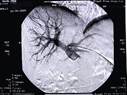
Portal vein embolization
Embolization of a portal vein (branch) is a radiological intervention procedure that is performed before planned resections of the liver parenchyma of a larger extent[1]. The goal of embolization of the portal vein leading the blood to the part of the liver is the gradual atrophy of this part of the liver and conversely the hypertrophy of the remaining parenchyma. This method is used to prevent the patient from getting into liver failure after extensive liver resection.[2]
Physiological basis of a procedure[edit | edit source]
A unique feature of the liver parenchyma is its regeneration. Cells can increase their number and size. Most nutrients are delivered to the liver via the portal vein (and not via the hepatic artery). Therefore, the portal vein is chosen for this procedure. The desired hypertrophy can be achieved in 2-4 weeks after the intervention[2]. 25% (in a healthy individual) to 40% of liver tissue (in affected liver - steatosis…) is sufficient to ensure the metabolic function of the liver[1]. The liver grows fastest during the first two weeks after exercise, then growth slows[2].
Performance indications[edit | edit source]
Patients planned for larger-scale liver resection are indicated for port embolization, where less functional liver tissue is thought to be left, and this may not be sufficient for metabolic needs (→ liver failure). These are extensive resections such as right / left lobectomy or extended lobectomy. These are patients with:
- primary liver cancer;
- liver metastases − synchronous and metachronous (especially colorectal cancer, breast cancer or Grawitz tumor);
- Klatskin's tumor (hilar tumor of extrahepatic bile ducts)).
Contraindications to the performance[edit | edit source]
he absolute contraindications coincide with the contraindications of surgical procedures. Relative contraindications are interventional radiological contraindications (bleeding disorders and the like). [2]
n metastatic liver disease, it is essential to rule out metastases in the rest of the liver prior to port embolization[1]. It has been proven that if there is any metase in the remaining parenchyma, then after the centralization of the portal blood, the metastasis will grow faster [2].
Prior examinations required[edit | edit source]
In addition to routine internal examinations, it is necessary[2]:
- CT (or MRI) − which maps the liver involvement (which liver segments are affected → these will need to be embolized, to rule out metastatic involvement of the remaining parenchyma);
- CT angio (or MRI) to clarify the portal vein before embolization;
- CT volumometry −to estimate the future residual capacity of the liver[1].
Custom performance[edit | edit source]
Embolization is performed percutaneously similar to PTC. Under X-ray control, a skin incision is made under local anesthesia in 10-11. intercostal space in the mid-axillary line and insert the needle deep into the liver parenchyma (porta is injected intrahepatically!). In order to detect that the needle is in the portal vein, a contrast agent is injected gradually as it moves. A thin wire is then inserted through the needle, followed by a metal cannula and a wire dilator. With the help of a contrast agent, the branches of the ports for the individual liver segments are clarified. The actual embolization is performed using gelatin foam, metal spirals or special substances. These are always applied centrally from the periphery and at the same time the balloon ensures that the embolization mass does not escape outside the required location.[2]
After the performance, coverage by various antibiotics is necessary.
Complications[edit | edit source]
- Pneumothorax;
- artery puncture;
- the formation of a fistula between the bile ducts and the venous system;
- other common surgical complications.
Links[edit | edit source]
Related articles[edit | edit source]
References[edit | edit source]
- ↑ a b c d VISOKAI, Vladimír. Chirurgická léčba u kolorektálního karcinomu jaterních metastáz [lecture for subject chirurgie-předstátnicová stáž, specialization general medicine, 1. LF UK Univerzita Karlova v Praze]. Praha. 2011-10-12.
- ↑ a b c d e f g VISOKAI, Vladimír – LIPSKÁ, Ludmila, et al. Recidiva kolorektálního karcinomu : Komplexní přístup z pohledu chirurga. 1. edition. Praha : Grada, 2009. 456 pp. pp. 174-180. ISBN 978-80-247-3026-4.

