Consequences of portal hypertension
Bleeding from oesophageal varices[edit | edit source]
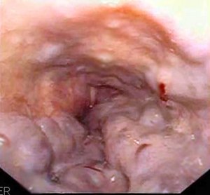
Endoscopy - bleeding from oesophageal varices.
Oesophageal varices are dilated veins in the submucosa of the distal esophagus. Bleeding from esophageal varices is a common complication of liver cirrhosis (30-60%).
Etiology and pathogenesis[edit | edit source]
The cause of varicose veins is long-lasting portal hypertension. Increased pressure leads to dilation of portocaval junctions, specifically between the left gastric vein (basin v. Portae), the oesophageal vein and the azygos vein (basin v. cava superior). There is a significant risk of bleeding from oesophageal area when the portal pressure rises above 12 mm Hg. Large varices (> 5 mm) and thin-walled varices bleed more often.
Clinical picture and diagnostics[edit | edit source]
A rupture of the varix leads to bleeding into the oesophagus, which irritates the stomach and usually results in bright red hematemesis. Subsequent melena is associated with the passage of blood through the digestive tract. With massive bleeding, signs of hemorrhagic shock begin to appear. Bleeding from esophageal varices is diagnosed by endoscopy.
Bleeding from oesophageal varices is the cause of death in half of patients with advanced liver cirrhosis.
Therapy[edit | edit source]
In the first line of hospitalisation in the ICU, ensuring central venous access and administration of erymass (RBCs) to compensate for blood loss. Subsequently, the bleeding is stopped endoscopically, by sclerosing, EVL. Due to the origin of varicose veins, sclerosing, operative removal of varicose veins, must be targeted at the cardiac veins. every 4 h, oesophageal variceal somatostatin (5 days).
With continued bleeding, endoscopic therapy is repeated, exceptionally, with unstoppable massive bleeding, the Sengstaken two-balloon nasogastric tube is introduced. The first balloon attaches to the stomach, where it compresses any bleeding, and the second balloon inflates in the oesophagus, compressing the varices. The balloons are left in situ for a maximum of 24 hours, otherwise ulceration will occur. Aspiration can be a complication.
Recurrence of bleeding is very common (up to 2 years 60–100% of patients) and β-blockers are used preventively (non-selective, in sufficient dose), from invasive solutions then repeated ligation/sclerosis of varices or TIPS/surgical portocaval joint surgery.
TIPS[edit | edit source]
TIPS (transjugular intrahepatic portosystemic shunt) works to prevent the formation and bleeding of oesophageal varices because it normalises portal hypertension. The design is three-step:
- hepatic vein cannulation via transjugular puncture,
- piercing of the liver parenchyma and probing of the v. portae branch,
- introduction of a self-expanding stent (communication between v. portae + v. hepatica).
Complications of performance are the emergence or worsening of encephalopathy, or clutch stenosis.
See the TIPS page for more information.
Prognosis[edit | edit source]
Bleeding has a high lethality and frequent recurrences (prevention of necessities).
Ascites[edit | edit source]
Ascites is an increased amount of free fluid in the abdominal cavity (the normal is up to 150 ml). Ascites is often accompanied by dyspepsia, flatulence, shortness of breath and difficulty moving. An umbilical hernia may occur due to increased intra-abdominal tension. A small amount of ascites may not cause any subjective difficulties.
Etiology[edit | edit source]
The most common cause of ascites is liver cirrhosis.
Other causes include:
- right heart failure;
- nephrotic syndrome;
- peritoneum carcinomatosis;
- pancreatitis;
- veno-occlusive disease, Budd-Chiari syndrome, portal vein thrombosis;
- TBC;
- ascites in dialysis, myxoedema, chlamydial infections.
Pathogenesis[edit | edit source]
In portal hypertension, the pressure in the hepatic sinusoids increases, leading to the penetration of albumin into the extravascular space. Fluid leakage into the hepatic interstitium increases. The fluid is drained by the lymphatic system, and if drainage is not sufficient, excess fluid escapes through the liver surface into the peritoneal cavity and ascites forms.
Classification[edit | edit source]
- small - detected on imaging methods
- medium-sized ascites detectable by thorough physical examination
- Large - clear on physical examination
Large ascites can be tense or non-tense. The abdomen is immobile when tense ascites occurs. Ascites that do not respond to the maximum possible diuretic treatment are referred to as refractory ascites.
Diagnostics[edit | edit source]
The basis of ascites diagnosis is physical examination and imaging. By physical examination, we can detect ascites in quantities of more than 2 litres. During the physical examination, we observe the abdomen at a level, or a smoothed navel or umbilical hernia. The palpable abdomen can be soft or hard during tension ascites. The tap is shortened. Undulation is present with a large amount of ascites (10 l or more).
Ultrasound examination also shows a small increase in peritoneal fluid. Ascites is imaged as an anechogenic fluid on ultrasound. Ascites is also imaged on other imaging methods (CT, magnetic resonance imaging). At the first occurrence of ascites or sudden deterioration of the patient with chronic liver disease, diagnostic ascites puncture and examination of laboratory parameters of ascites fluid are indicated in order to exclude spontaneous bacterial peritonitis and determine serum-ascites albumin gradient (SAAG> 11 g / l → PH).
Therapy[edit | edit source]
- Treatment of the cause of ascites - alcohol abstinence in patients with alcoholic liver cirrhosis, antiviral treatment in patients with liver cirrhosis based on chronic viral hepatitis B or C, etc.,
- limiting the salt to 3 grams per day; fluids usually do not need to be severely restricted (fluid restriction is recommended for hyponatraemia <120 mmol / l),
- exclusion of nephrotoxic medication (aminoglycosides, antiphlogistics),
- diuretics - spironolactone and furosemide in individually determined doses, usually in a ratio of 100: 40 mg per day and in maximum doses up to 400 mg / day spironolactone and 160 mg / day furosemide,
- paracentesis (ascites discharge) - after that i.v. albumin = prevention of hypovolemia (renal circulatory disorder to shock),
- TIPS (transjugular intrahepatic portosystemic shunt) - prevents reacculation of ascites, indicated in refractory ascites,
- surgical introduction of an artificial peritoneovenous junction (Le Veenova, Denver), which drains ascites directly into the central vein - this method is rarely used.
Spontaneous bacterial peritonitis[edit | edit source]
Spontaneous bacterial peritonitis is a bacterial infection of the ascites without a detectable, surgically treatable source of infection. This is a common complication of ascites (30%) of cirrhotic origin.
Etiology and pathogenesis[edit | edit source]
- The source of the infection is probably the intestine - the infection passes through the intact intestinal wall through translocations,
- more susceptible ascites with low opsonin activity,
- originator hl. facultatively anaerobic gram-negative intestinal bacteria: E. coli, Klebsiella, Enterobacter, Proteus.
Clinical picture[edit | edit source]
- Symptoms are variable and mostly insignificant,
- infection can manifest itself only as increasing accumulation of ascites + failure of diuretic treatment or hepatic impairment,
- subfebrile + diffuse abdominal pain,
- often occurs after bleeding from oesophageal varices,
- Has a lethality of about 30% when untreated.
Diagnostics[edit | edit source]
- Diagnostic paracentesis with ascites examination: culture, leukocytes> 0.4 x 10⁹ / l → start treatment.
Therapy[edit | edit source]
- cephalosporins III. generation (cefotaxime 2 g every 8 hours), albumin (prevention of hypovolemia - hepatorenal syndrome),
- selective intestinal decontamination with non-absorbable ATB (norfloxacin 400 g) - prevention.
Prognosis[edit | edit source]
- Poor (recurrences, worsening of liver + renal function).
Hepatic encephalopathy[edit | edit source]
Hepatic encephalopathy is a set of reversible neuropsychiatric symptoms. It occurs in acute liver failure, in which liver cells die and thus liver detoxification function is impaired. Hepatic encephalopathy also occurs in chronic liver disease, most commonly in cirrhosis of the liver. Hepatic encephalopathy is caused by an increased concentration of substances normally metabolized by the liver, which act in the CNS to inhibit nerve transmission. It is mainly a change in transmission at GABA receptors. These substances are, for example, ammonia, neurosteroids, glutamine, phenols, mercaptans.
Clinical symptoms[edit | edit source]
There are 4 stages:
- In the first stage, the patient is slightly confused, sleep and behavioral disorders occur.
- In the second stage, personality and thinking disorders are present.
- In the third stage, the patient experiences somnolence and disorientation.
- The fourth and final stage is defined by coma.
Specific symptoms are flapping tremor and foetor hepaticus.
Diagnostics[edit | edit source]
It includes a laboratory test, in which we look for increased ammonia, which is a high level of ammonia in the blood.
We also perform auxiliary examinations:
- number connection test (latency when connecting scattered numbers according to a sequence),
- structural apraxia (eg unable to draw a star),
- EEG, evoked potentials (ocular, auditory, cognitive),
- CT or MRI of the brain are important in differential diagnosis.
Differential diagnostics[edit | edit source]
- Alcoholism (withdrawal syndrome, delirium tremens),
- Wilson's disease.
Treatment[edit | edit source]
The ideal treatment is liver transplantation, because after its implementation, changes in the body are usually fully reversible. It is also possible to limit protein intake and administer lactulose or lactitol, which serve to induce osmotic diarrhea. ATBs are also given against the intestinal microflora, which reduces the amount of ammonia produced by the intestinal bacteria. For example, rifaximin is used, which acts only on the intestinal microflora and has no systemic effects and associated systemic side effects.
Hepatorenal syndrome[edit | edit source]
Hepatorenal syndrome is a functional kidney failure in liver disease with portal hypertension. It occurs almost exclusively in patients with ascites.
Etiology and pathogenesis[edit | edit source]
The basis is systemic circulatory changes in portal hypertension.
- Renal arterial vasoconstriction (with cortical hypoperfusion) + renal impairment,
- the basis is systemic circulatory changes in portal hypertension (↓ peripheral vascular resistance, central hypovolemia, sympathetic activation).
Clinical picture[edit | edit source]
- Type I - rapidly progressing, 2x ↑ serum creatinine within 2 weeks, very poor prognosis,
- Type II - slowly progressing, renal insufficiency occurs slowly + condition relatively stabilised.
Diagnostics[edit | edit source]
There is no specific test that can diagnose hepatorenal syndrome. Glomerular filtration is usually <0.66 ml / s (40 ml / min), serum creatinine> 135 μmol / l, sodium in urine <10 mmol / l, urinary osmolality> plasma.
Differential diagnostics[edit | edit source]
- Organic kidney damage (ATN, etc.).
Therapy[edit | edit source]
- Exclusion: nephrotoxic drugs, diuretics, non-steroidal anti-inflammatory drugs,
- treat bacterial infection, eliminate bleeding into the gastrointestinal tract,
- correction of hypovolemia (albumin, terlipressin),
- TIPS (days to weeks apart),
- liver transplantation.
Links[edit | edit source]
Related articles[edit | edit source]
External links[edit | edit source]
References[edit | edit source]
- DÍTĚ, P., et al. Vnitřní lékařství. 2. vydání. Praha : Galén, 2007. ISBN 978-80-7262-496-6.
- PASTOR, Jan. Langenbeck's medical web page [online]. [cit. 2010]. <https://langenbeck.webs.com/>.
- ↑ POVÝŠIL, Ctibor. Speciální patologie. - vydání. Galén, 2007. 430 s. s. 145. ISBN 9788072624942.


