Preparation of histological specimen
A histological specimen is prepared and processed using a histological technique so that we can examine the microscopic structures of the tissues, establish a diagnosis and rule out possible pathologies. For further work with histological samples, it is absolutely necessary to fix, embed, cut, stain and mount them in a glass slide.
Fixace[edit | edit source]
After removing the tissue for histological examination, the sample must be quickly fixed, i.e. preserved against autolysis (the action of its own enzymes, which can, for example, make it impossible to stain the sample). The treatment of the sample consists of immersing the tissue in a stabilizing (fixing) agent or fixative, which cause a gentle denaturation of the enzyme proteins.
Fixation prevents deterioration of the sample. There are three main requirements for successful fixation: gentleness (good preservation of the structure of cells and tissues), speed of penetration into the sample and enabling its further processing. Of course, fixing agents have their advantages and disadvantages. Some gently denature proteins, while others, on the contrary, cause them to clump together. That is why fixing compounds have been developed that have the most favorable properties.
- Baker's fixative – contains 10% formaldehyde, calcium chloride and water. It does not dissolve fats, therefore it is suitable for lipid detection.
- Bouin solution – contains picric acid, formaldehyde, acetic acid and water. It is suitable for the detection of polysaccharides, on the contrary, it is not suitable for the detection of lipids and proteins.
- Zenker's fixative – contains mercuric chloride, potassium dichromate, potassium sulfate, acetic acid and water. It is an exception among fixative fluids because it does not contain formaldehyde.
Another frequently used fixative is formaldehyde, usually in an aqueous solution at a concentration of around 4% (a 40% aqueous solution of formaldehyde is traditionally referred to as "formalin"). It is used for its thriftiness and low price.
In electron microscopy, osmium oxide is used, mainly due to the increase in the contrast of structures and its gentle properties (it penetrates the tissue very slowly).
Fixation can be not only chemical, but also physical - e.g. quick freezing of the tissue, freezing-drying method, or microwave radiation.
Embedding tissue[edit | edit source]
Fixed samples must be cut into thin sections. To be able to cut the tissue on the microtome, it must be embedded in the mounting medium.
The most common medium is paraffin. Not suitable for hard tissues, e.g. teeth. When embedding for electron microscopy purposes, the tissues are impregnated with artificial resins.
Before embedding, the samples are dehydrated and cleared. Water is removed by alcohol baths of increasing concentration. It usually starts with 70% and ends with 100% ethanol, however, the initial concentration is chosen according to the water content in the tissue. After that, the bath is replaced with a solvent that is mixed with the embedding medium (benzene, xylene, toluene are used for embedding in paraffin). We already call this phase clearing, during which the tissue saturated with the solvent becomes translucent.
The cleared tissue is transferred to paraffin melted at a boundary temperature of 52-60 °C[1] when the paraffin is still liquid and the tissue is not damaged. The solvent is removed from the tissue in three baths and is subsequently replaced with paraffin. It is then followed by embedding in high-quality paraffin and solidification (usually at room temperature).
Sectioning tissue[edit | edit source]
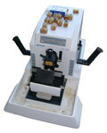
The solidified tissue block is sliced (trimmed) to get rid of excess embedding medium. In order to properly examine the sample under the microscope, it is important to cut it into very thin sections. Tissues are cut on so-called microtomes (sled or rotary). Samples intended for histochemical methods (often embedded in gelatin) are cut on a cryostat (freezing microtome). Samples intended for electron microscopy are cut on ultramicrotomes.
Sections are cut to an adjustable thickness – 1–10 µm (ideally around 6–8 µm). Ultramicrotomes cut around 0.02–0.1 µm.[2]
The sections are then transferred to a warm water surface where they are so-called stretched and then transferred to a glass slide. Slides with sections are placed in a thermostat to get rid of water.
Staining sections[edit | edit source]
Most histological structures are colorless, so to be able to recognize individual tissue formations, the preparations usually have to be stained.
Prepared cuts are most often infiltrated with paraffin, which is immiscible with water. The stains used are usually dissolved in water or alcohol, so it is necessary to remove the paraffin from the tissue. The so-called deparaffinization is carried out in xylene, followed by a descending series of alcohols (so-called water-down) with decreasing concentrations (96%, 80%, 70%). After this procedure, the sample is ready for staining.
Most histological stains behave as acidic or basic substances. According to chemical affinity, we divide the structures into basophilic and acidophilic. Basophilic structures (acids) are stained with basic dyes - e.g. hematoxylin. Typically, this is, for example, a nucleus with DNA. Acidophilic structures (bases) are stained with acid dyes - e.g. eosin - which is why the term eosinophilic is also sometimes used. Cytoplasm is typically acidophilic.
The most commonly used coloring mixture is:
- hematoxylin and eosin (H&E stain) – hematoxylin stains blue-violet, eosin pink-red. This is clear staining.
Special stains are also used , e.g.:
- Masson's trichromes – yellow, blue and green, distinguish collagen fiber and muscle;
- alcian blue – highlights glycosaminoglycans;
- impregnation with silver – staining of reticular fibers, neurons and axons, glia.
Mounting of section[edit | edit source]
The last stage of preparing the specimen is gluing the coverslip. Before gluing the slide, a mounting medium is dripped onto this cut, previously often used Canadian balsam, now artificial epoxy resin (insoluble in water). If the specimen cannot be mounted using a medium with an organic solvent, a water-based medium (e.g. glycerin) is used.
Specimen intended for electron microscopy are placed loosely on metal grids (electrons cannot penetrate the glass).
Links[edit | edit source]
Related articles[edit | edit source]
- Blood smear
- Preparation of samples for histological examination by light and electron microscopy
- Biopsy
- Staining in light microscopy
Reference[edit | edit source]
References[edit | edit source]
- JUNQUIERA, L. Carlos – CARNEIRO, José – KELLEY, Robert O. Základy histologie. 1. edition. Jinočany : H & H, 1997. 502 pp. ISBN 80-85787-37-7.
- JIRKOVSKÁ, Marie. Histologická technika. 1. edition. 2006. ISBN 80-7262-263-4.
- MESCHER, Anthony. Junqueira's Basic Histology: Text and Atlas, Fourteenth Edition. - edition. McGraw-Hill Education, 2015. 1136 pp. ISBN 9780071842709.


